Advanced in vitro Modeling of Human iPSC-derived Neuronal Mono- and Co-cultures with Microglia: Optimization Using Growth Factors and Live-Cell Analysis
Human induced pluripotent stem cell (iPSC) derived models are increasingly being used for in vitro studies of the human central nervous system. These models enable the generation of specific neurons and support cells and provide a more translational system for investigating human development and disease. Growth factors and cytokines are critical in these models for cell growth, differentiation, and survival, but their study is complicated by factors like unknown commercial media compositions and sensitivity to handling. To successfully use these advanced models, robust culture conditions and technology approaches to non-invasively optimise, visualise, and kinetically quantify these complex models are required.
In this application note, the Sartorius team examines the efficacy of Sartorius Research Use Only (RUO) Growth Factors and Cytokines for robustly maintaining iPSC-derived neuronal and microglial models in healthy and diseased states and demonstrate how the Incucyte® Live-Cell Analysis System facilitates their characterisation through non-invasive, continuous monitoring of culture health, morphology, marker expression, and function.
In this application note, you will discover:
- An integrated solution for the visualisation and quantification of long-term changes in the health, morphology, function, and marker expression in ioMicroglia and ioGlutamatergic Neuron cell models.
- Methods for the robust co-culture of ioGlutamatergic Neuron models in healthy and diseased states with ioMicroglia utilizing Sartorius’ Research Use Only (RUO) Growth Factors and Cytokines.
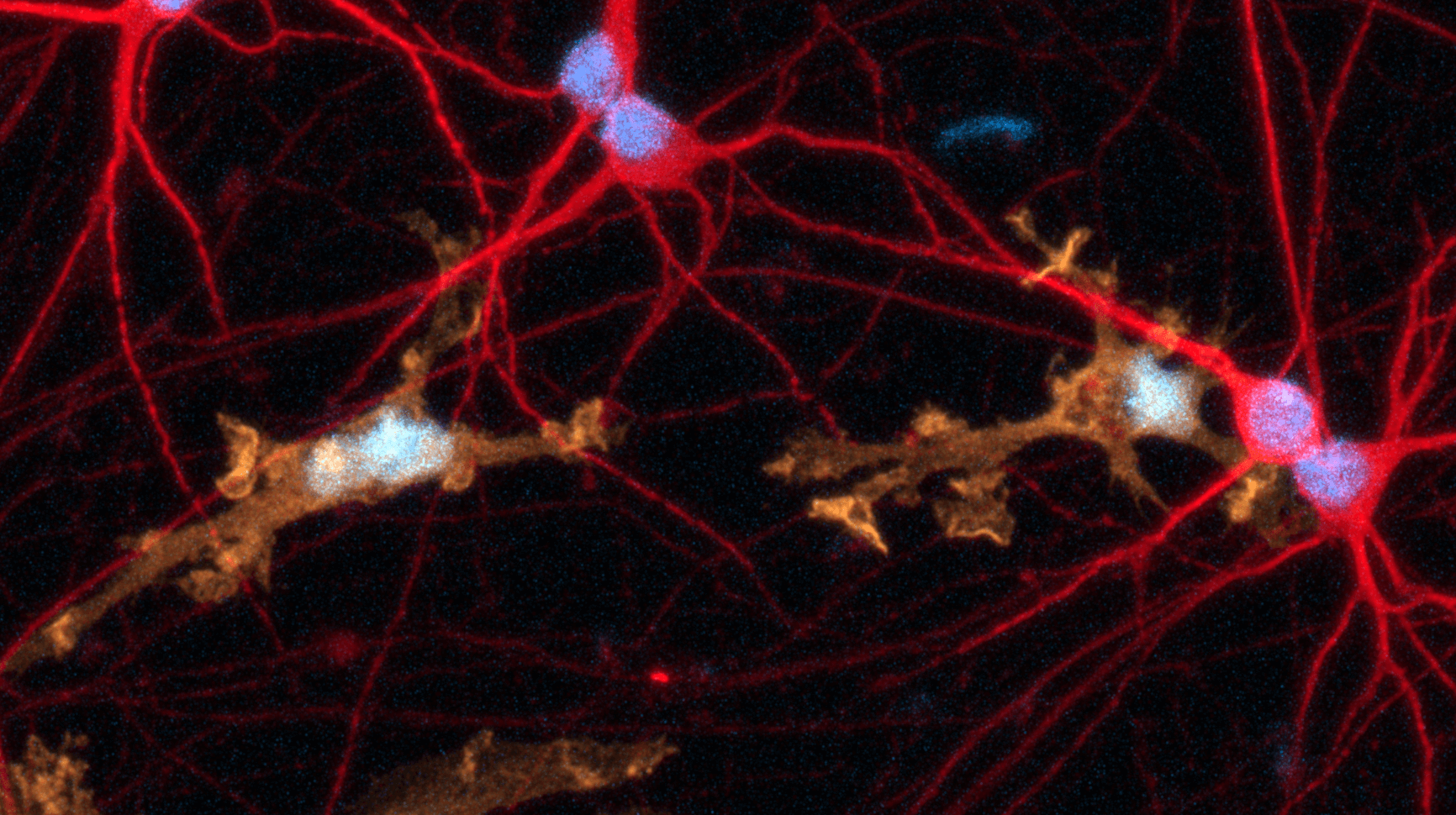
- A case study on the response of ioMicroglia following treatment with pro-inflammatory stimuli showing that these cells exhibit stellate morphology indicative of microglial activation.
- A case study on the quantification of dynamic changes of Huntington’s disease model ioGlutamatergic Neurons compared to a genetically matched control, monitoring their neurite length and branching. Calcium imaging assays revealed that co-culture of healthy and disease model ioGlutamatergic Neurons with ioMicroglia synchronised calcium bursts, and that these bursts were markedly different in the disease model.
This application note was originally published by Sartorius
Sartorius
2024