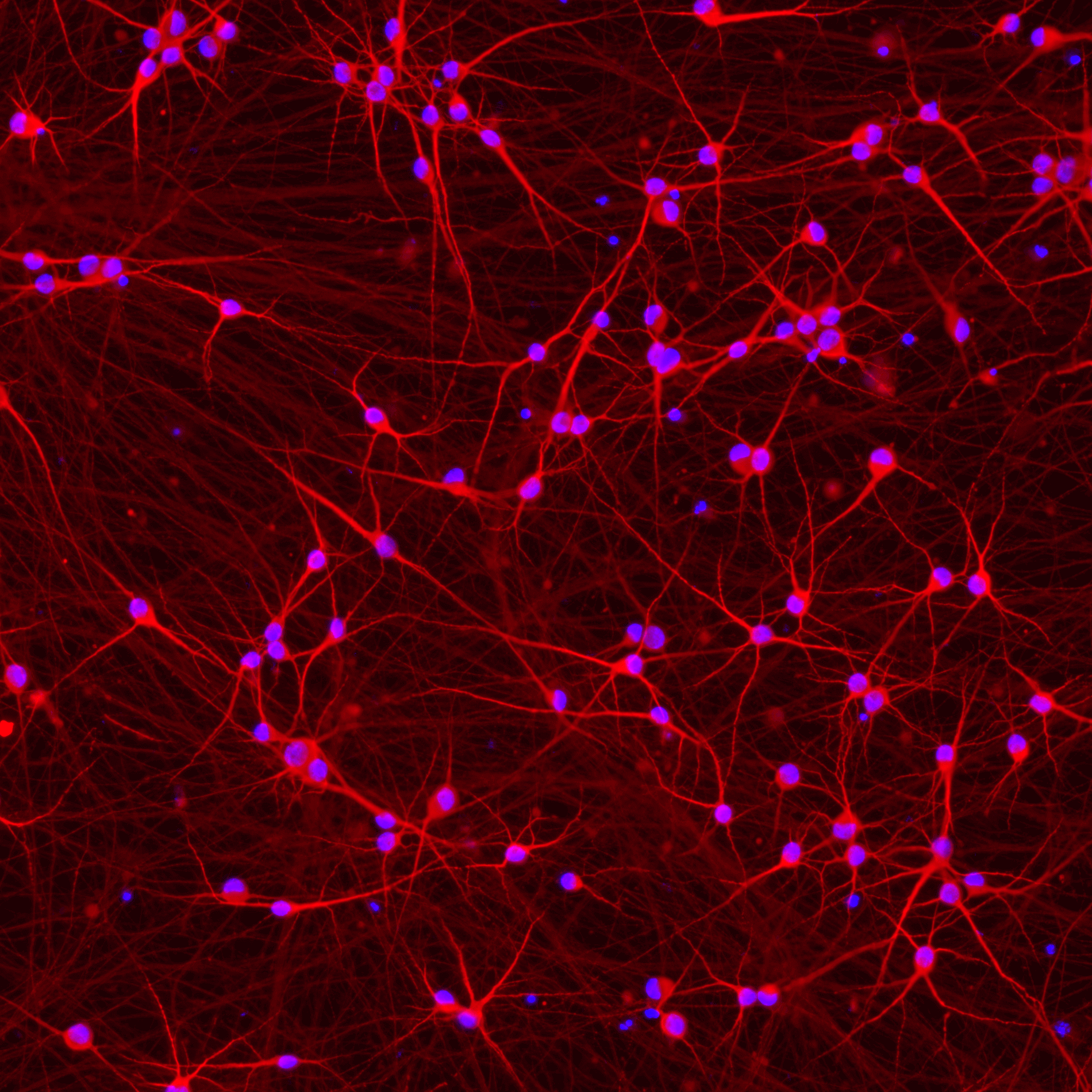
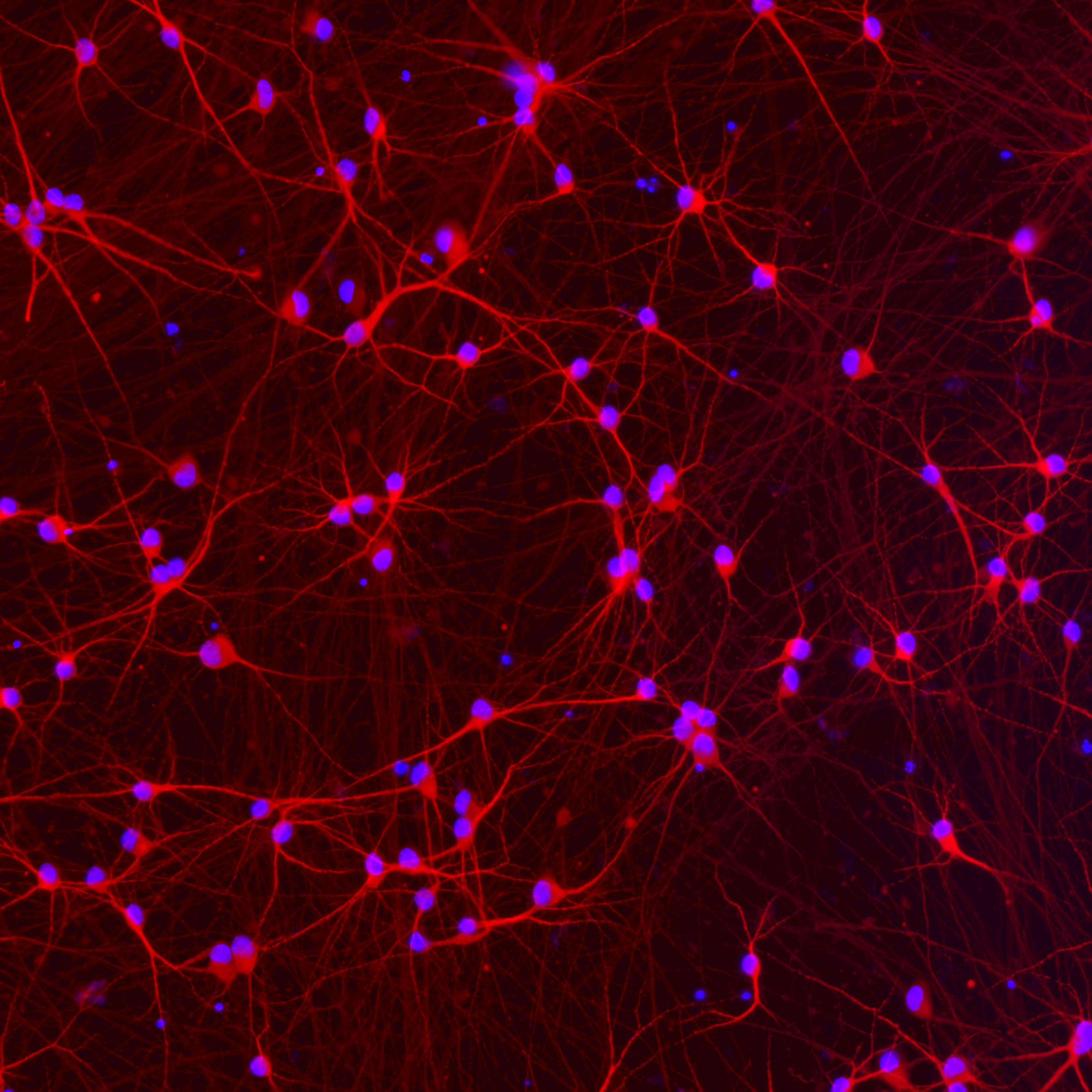
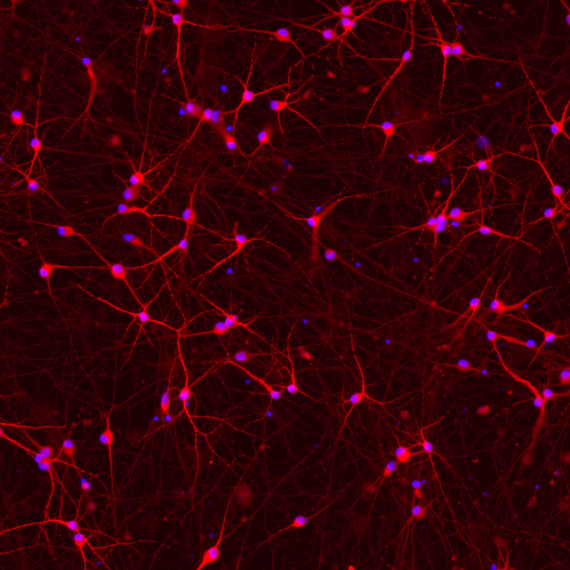
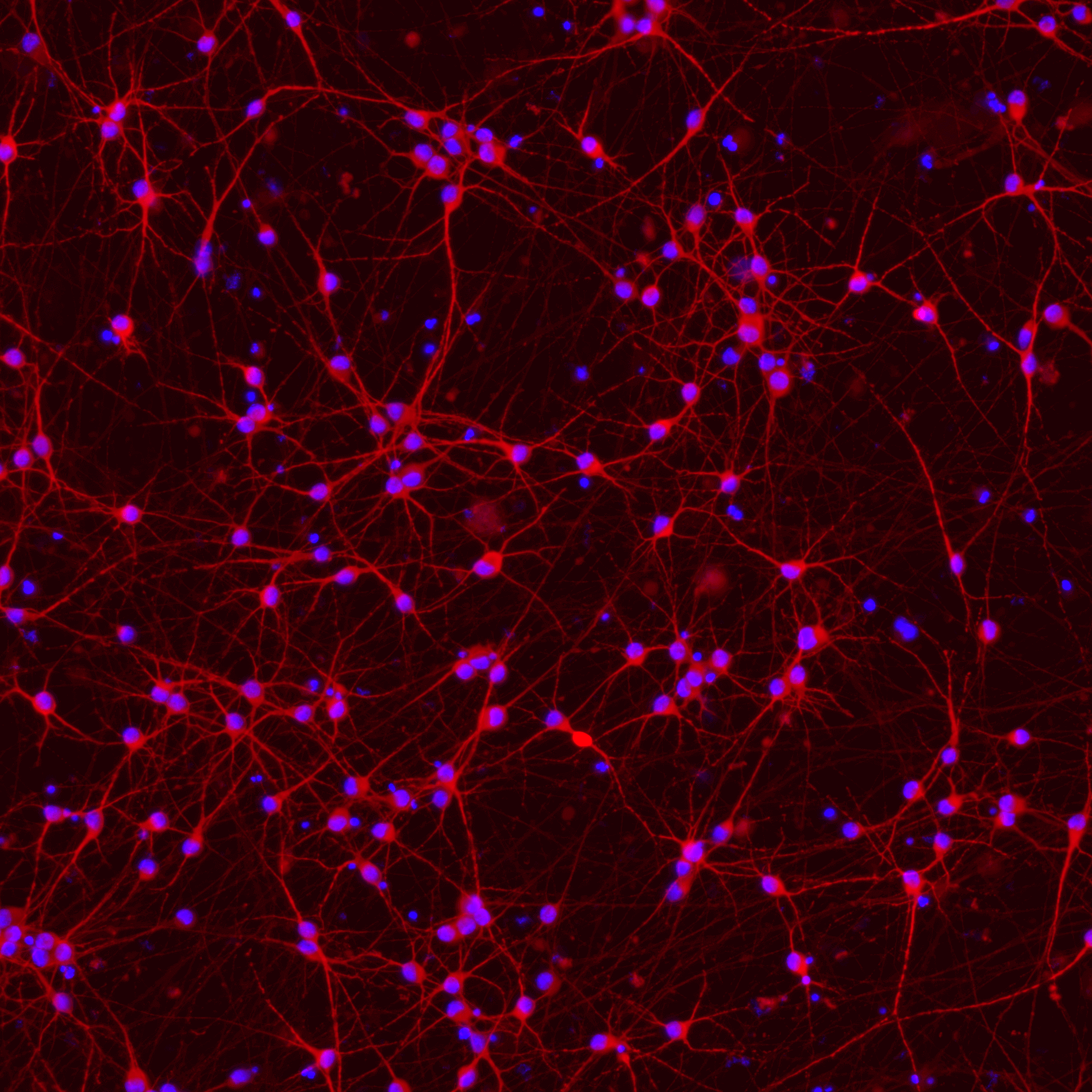
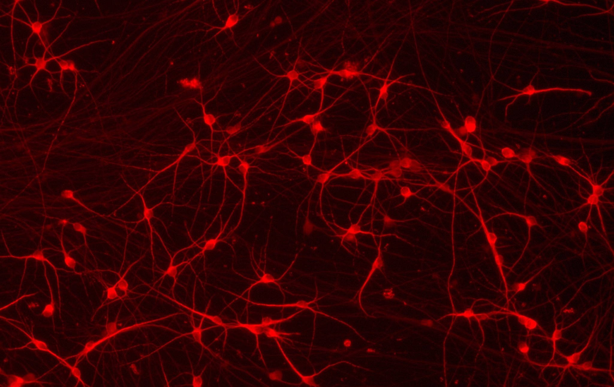
Human iPSC-derived Alzheimer's disease model in glutamatergic neurons
bit.bio Increased ratio of amyloid beta 42:40 in glutamatergic neurons APP V717I London mutation
bit.bio Staining of glutamatergic neuron markers beta3 tubulin, MAP2 and VGLUT2 in the APP V717I hom disease model
bit.bio Brightfield imaging of iPSC derived neurons APP V717I hom disease model shows glutamatergic neurons morphology
bit.bio Gene expression of neuronal markers in glutamatergic neurons APP V717I Alzheimer's disease model
bit.bio Gene expression of APP in glutamatergic neurons APP V717I Alzheimer's disease model
bit.bio Glutamatergic neurons APP V717I hom are suitable for culture in 96-well and 384-well plates for screening applications

cat no | io1063

ioGlutamatergic Neurons APP V717I/V717I

Human iPSC-derived Alzheimer's disease model

  • Cryopreserved human iPSC-derived cells powered by opti-ox that are ready for experiments in days

  • Functional excitatory neurons engineered with the APP London mutation for Alzheimer's disease research

  • Disease-related phenotype demonstrated by increased ratio of A𝛽42:40 peptides vs. wild-type control

Place your order

Human iPSC-derived Alzheimer's disease model in glutamatergic neurons

Human iPSC-derived Alzheimer's disease model

bit.bio Increased ratio of amyloid beta 42:40 in glutamatergic neurons APP V717I London mutation

Increased ratio of A𝛽42:40 seen in ioGlutamatergic Neurons APP V717I (London), as observed in Alzheimer’s disease

ioGlutamatergic Neurons APP V717I disease model cells show increased production of A𝛽38 and A𝛽42 peptides (involved in the amyloidogenic pathway), with no difference seen for A𝛽40 (A). This results in an increased ratio of A𝛽42:40 and no change in the A𝛽42:38 ratio (B).

  • ioGlutamatergic Neurons wild type (WT), APP V717I/WT (CL35, io1067S), and APP V717I/V717I (CL27, io1063S), were seeded at 30,000 cells/cm2 in 24-well plates and cultured for 30 days according to the user manual. Supernatant was collected at days 10, 20, and 30.
  • Levels of A𝛽38, A𝛽40 and A𝛽42 peptides were quantified using the V-PLEX A𝛽 Peptide Panel 1 (6E10) Kit (MSD K15200E-1).
  • Concentrations of A𝛽38, A𝛽40, A𝛽42 were normalised to the calculated total number of cells per well.
  • Data were obtained from two independent experiments and are shown as mean ± SEM. Data were analysed statistically (at days 20 and 30) using Student’s t-tests comparing each clone to the wild type.
    * p<0.05 ** p<0.01 ***p<0.001
bit.bio Staining of glutamatergic neuron markers beta3 tubulin, MAP2 and VGLUT2 in the APP V717I hom disease model

ioGlutamatergic Neurons APP V717I/V717I express neuron-specific markers comparably to the wild type control

Immunofluorescent staining on post-revival day 11 demonstrates similar homogenous expression of pan-neuronal proteins MAP2 and TUBB3 (upper panel) and glutamatergic neuron-specific transporter VGLUT2 (lower panel) in ioGlutamatergic Neurons APP V717I/V717I (CL27) compared to the genetically matched control. 100X magnification.

bit.bio Brightfield imaging of iPSC derived neurons APP V717I hom disease model shows glutamatergic neurons morphology

ioGlutamatergic Neurons APP V717I/V717I form structural neuronal networks by day 11

ioGlutamatergic Neurons APP V717I/V717I mature rapidly, show glutamatergic neuron morphology and form structural neuronal networks over 11 days, highly similar to the genetically matched control. Day 1 to 11 post thaw; 100X magnification.

bit.bio Gene expression of neuronal markers in glutamatergic neurons APP V717I Alzheimer's disease model

ioGlutamatergic Neurons APP V717I/V717I demonstrate gene expression of neuronal-specific and glutamatergic-specific markers following deterministic cell programming

Gene expression analysis demonstrates that ioGlutamatergic Neurons APP V717I/V717I (CL27, io1063) and wild type ioGlutamatergic Neurons (WT Control) lack the expression of pluripotency markers (NANOG and OCT4) at day 11, while robustly expressing pan-neuronal (TUBB3 and SYP) and glutamatergic-specific (VGLUT1 and VGLUT2) markers, as well as the glutamate receptor GRIA4. Gene expression levels were assessed by RT-qPCR (data normalised to HMBS; cDNA samples of the parental human iPSC line (hiPSC) were included as reference). Data represents day 11 post-revival samples, n=2 replicates.

bit.bio Gene expression of APP in glutamatergic neurons APP V717I Alzheimer's disease model

Disease-related APP is expressed in ioGlutamatergic Neurons APP V717I/V717I following deterministic programming

RT-qPCR analysis demonstrates expression of the APP gene in both wild type ioGlutamatergic Neurons (WT Control) and ioGlutamatergic Neurons APP V717I/V717I at day 11 post-revival. Data normalised to HMBS, n=2 replicates.

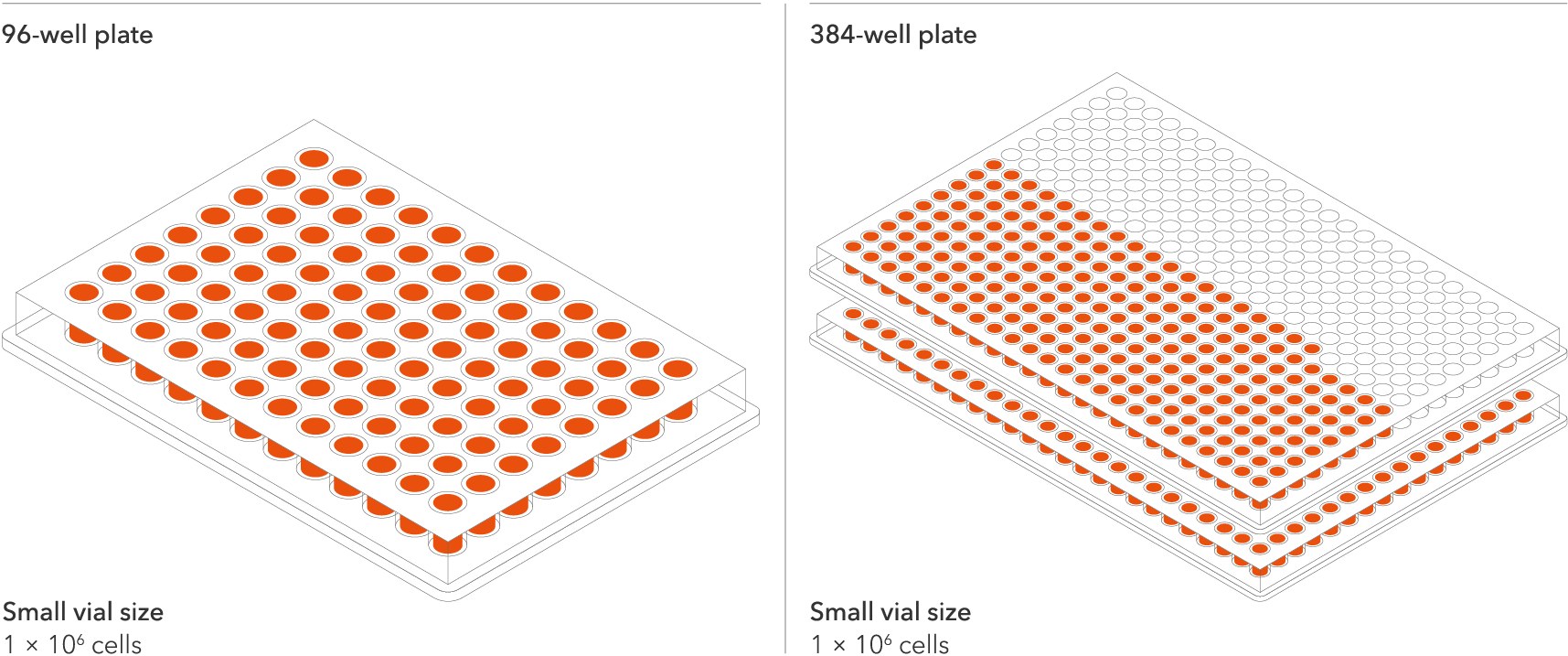
bit.bio Glutamatergic neurons APP V717I hom are suitable for culture in 96-well and 384-well plates for screening applications

Industry leading seeding density

The recommended minimum seeding density is 30,000 cells/cm2, compared to up to 250,000 cells/cm2 for other similar products on the market. One small vial can plate a minimum of 0.7 x 24-well plate, 1 x 96-well plate, or 1.5 x 384-well plates. This means every vial goes further, enabling more experimental conditions and more repeats, resulting in more confidence in the data.

Vial limit exceeded

A maximum number of 20 vials applies. If you would like to order more than 20 vials, please contact us at orders@bit.bio.

Human iPSC-derived Alzheimer's disease model

A rapidly maturing, physiologically relevant, functional system for investigating the role of the APP London mutation in early-onset Alzheimer's disease (AD).  This in vitro disease cell model recapitulates an increased ratio of amyloid beta peptides A𝛽42:40, as observed in AD.

ioGlutamatergic Neurons APP V717I/V717I are opti‑ox deterministically programmed excitatory neurons carrying a genetically engineered homozygous V717I mutation in the APP gene encoding amyloid precursor protein.

This disease model is part of an Alzheimer's disease panel of human iPSC-derived cells that can be incorporated into translational research and drug discovery workflows. Two additional clones for the APP V717I hom mutation are available for scientists who wish to repeat their experiments in multiple independent clones, please enquire. All disease models are genetically matched to the wild-type control, ioGlutamatergic Neurons. Additional mutations in the AD panel include heterozygous APP V717I, and heterozygous and homozygous APP KM670/671NL and PSEN1 M146L, alongside AD-relevant mutations in ioGABAergic Neurons and ioMicroglia.

Benchtop benefits

Phenotype of increased ratio of A𝛽42:40

Disease related phenotype

Increased ratio of A𝛽42:40 peptides compared to the genetically matched control, measured by immunoassay.

Compare disease model with wild type control

Make True Comparisons

Pair the Alzheimer's disease model cells with the wild type glutamatergic neurons to investigate the impact of the APP missense mutation.

Glutamatergic neurons are quick to culture

Quick

The disease model cells and genetically matched control are experiment ready as early as 2 days post revival, and form structural neuronal networks at 11 days.

Cells arrive ready to plate


ioGlutamatergic_Neurons_and_disease_models_timeline

ioGlutamatergic Neurons APP V717I/V717I are delivered in a cryopreserved format and are programmed to mature rapidly upon revival in the recommended media. The protocol for the generation of these cells is a two-phase process: Phase 1, Stabilisation for 4 days; Phase 2, Maintenance, during which the neurons mature. Phases 1 and 2 after revival of cells are carried out by the customer.

Product specifications

Starting material

Human iPSC line

Karyotype

Normal (46, XY)

Seeding compatibility

6, 12, 24, 96 & 384 well plates

Shipping info

Dry ice

Donor

Caucasian adult male, age 55-60 years old (skin fibroblast),
Genotype APOE 3/4

Vial size

Small: >1 x 10⁶ viable cells, Evaluation pack*: 3 small vials of >1 x 10⁶ viable cells

Quality control

Sterility, protein expression (ICC), gene expression (RT-qPCR) and genotype validation (Sanger sequencing)

Differentiation method

opti-ox deterministic cell programming

Recommended seeding density

30,000 cells/cm²

User storage

LN2 or -150°C

Format

Cryopreserved cells

Genetic modification

Homozygous V717I missense mutation in the APP gene

Applications

Alzheimer's disease research
Drug discovery and development
Disease modelling

Available clones

io1063 | APP V717I/V717I (CL27)
io1064 | APP V717I/V717I (CL84)
io1065 | APP V717I/V717I (CL96)

Product use

ioCells are for research use only

* Evaluation packs are intended for first-time users, or for existing users testing a new cell type or derivative. A user can request multiple evaluation packs as long as each one is for a different product, with only one pack allowed per product.

Technical data

Disease related phenotype

Increased ratio of A𝛽42:40 seen in ioGlutamatergic Neurons APP V717I (London), as observed in Alzheimer’s disease

bit.bio Increased ratio of amyloid beta 42:40 in glutamatergic neurons APP V717I London mutation

ioGlutamatergic Neurons APP V717I disease model cells show increased production of A𝛽38 and A𝛽42 peptides (involved in the amyloidogenic pathway), with no difference seen for A𝛽40 (A). This results in an increased ratio of A𝛽42:40 and no change in the A𝛽42:38 ratio (B).

  • ioGlutamatergic Neurons wild type (WT), APP V717I/WT (CL35, io1067S), and APP V717I/V717I (CL27, io1063S), were seeded at 30,000 cells/cm2 in 24-well plates and cultured for 30 days according to the user manual. Supernatant was collected at days 10, 20, and 30.
  • Levels of A𝛽38, A𝛽40 and A𝛽42 peptides were quantified using the V-PLEX A𝛽 Peptide Panel 1 (6E10) Kit (MSD K15200E-1).
  • Concentrations of A𝛽38, A𝛽40, A𝛽42 were normalised to the calculated total number of cells per well.
  • Data were obtained from two independent experiments and are shown as mean ± SEM. Data were analysed statistically (at days 20 and 30) using Student’s t-tests comparing each clone to the wild type.
    * p<0.05 ** p<0.01 ***p<0.001

Highly characterised and defined

ioGlutamatergic Neurons APP V717I/V717I express neuron-specific markers comparably to the wild type control

bit.bio Staining of glutamatergic neuron markers beta3 tubulin, MAP2 and VGLUT2 in the APP V717I hom disease model

Immunofluorescent staining on post-revival day 11 demonstrates similar homogenous expression of pan-neuronal proteins MAP2 and TUBB3 (upper panel) and glutamatergic neuron-specific transporter VGLUT2 (lower panel) in ioGlutamatergic Neurons APP V717I/V717I (CL27) compared to the genetically matched control. 100X magnification.

ioGlutamatergic Neurons APP V717I/V717I form structural neuronal networks by day 11

bit.bio Brightfield imaging of iPSC derived neurons APP V717I hom disease model shows glutamatergic neurons morphology

ioGlutamatergic Neurons APP V717I/V717I mature rapidly, show glutamatergic neuron morphology and form structural neuronal networks over 11 days, highly similar to the genetically matched control. Day 1 to 11 post thaw; 100X magnification.

ioGlutamatergic Neurons APP V717I/V717I demonstrate gene expression of neuronal-specific and glutamatergic-specific markers following deterministic cell programming

bit.bio Gene expression of neuronal markers in glutamatergic neurons APP V717I Alzheimer's disease model

Gene expression analysis demonstrates that ioGlutamatergic Neurons APP V717I/V717I (CL27, io1063) and wild type ioGlutamatergic Neurons (WT Control) lack the expression of pluripotency markers (NANOG and OCT4) at day 11, while robustly expressing pan-neuronal (TUBB3 and SYP) and glutamatergic-specific (VGLUT1 and VGLUT2) markers, as well as the glutamate receptor GRIA4. Gene expression levels were assessed by RT-qPCR (data normalised to HMBS; cDNA samples of the parental human iPSC line (hiPSC) were included as reference). Data represents day 11 post-revival samples, n=2 replicates.

Disease-related APP is expressed in ioGlutamatergic Neurons APP V717I/V717I following deterministic programming

bit.bio Gene expression of APP in glutamatergic neurons APP V717I Alzheimer's disease model

RT-qPCR analysis demonstrates expression of the APP gene in both wild type ioGlutamatergic Neurons (WT Control) and ioGlutamatergic Neurons APP V717I/V717I at day 11 post-revival. Data normalised to HMBS, n=2 replicates.

Industry leading seeding density

bit.bio Glutamatergic neurons APP V717I hom are suitable for culture in 96-well and 384-well plates for screening applications

The recommended minimum seeding density is 30,000 cells/cm2, compared to up to 250,000 cells/cm2 for other similar products on the market. One small vial can plate a minimum of 0.7 x 24-well plate, 1 x 96-well plate, or 1.5 x 384-well plates. This means every vial goes further, enabling more experimental conditions and more repeats, resulting in more confidence in the data.

Technical data

Increased ratio of A𝛽42:40

Increased ratio of A𝛽42:40 seen in ioGlutamatergic Neurons APP V717I (London), as observed in Alzheimer’s disease

bit.bio-Glutamatergic-Neurons-APP-V717I-Abeta42-40-ratio-quantification

ioGlutamatergic Neurons APP V717I disease model cells show increased production of A𝛽38 and A𝛽42 peptides (involved in the amyloidogenic pathway), with no difference seen for A𝛽40 (A). This results in an increased ratio of A𝛽42:40 and no change in the A𝛽42:38 ratio (B).

  • ioGlutamatergic Neurons wild type (WT), APP V717I/WT (CL35, io1067S), and APP V717I/V717I (CL27, io1063S), were seeded at 30,000 cells/cm2 in 24-well plates and cultured for 30 days according to the user manual. Supernatant was collected at days 10, 20, and 30.
  • Levels of A𝛽38, A𝛽40 and A𝛽42 peptides were quantified using the V-PLEX A𝛽 Peptide Panel 1 (6E10) Kit (MSD K15200E-1).
  • Concentrations of A𝛽38, A𝛽40, A𝛽42 were normalised to the calculated total number of cells per well.
  • Data were obtained from two independent experiments and are shown as mean ± SEM. Data were analysed statistically (at days 20 and 30) using Student’s t-tests comparing each clone to the wild type.
    * p<0.05 ** p<0.01 ***p<0.001

How to culture ioGlutamatergic Neurons

In this video, our scientist will take you through the step-by-step process of how to thaw, seed and culture ioGlutamatergic Neurons.

Product resources

(LinkedValues: [{hs_name=Modelling neurodevelopment | Investigating the impact of maternal immune activation on neurodevelopment using human iPSC-derived cells, hs_id=161968263519, hs_path=modelling-neurodevelopment-webinar-2022, button_label=Explore ioGlutamatergic Neurons, button_link=https://www.bit.bio/products/nerve-cells/glutamatergic-neurons-wild-type-io1001, type={value=Webinar, label=Webinar}, thumbnail={alt_text=, width=1200, url=https://14527135.fs1.hubspotusercontent-na1.net/hubfs/14527135/BitBio2022/product-page/Colour%20webinar%20with%2060x%20Map2-com.jpg, height=732}, year={value=2022, label=2022}, summary=<p><span>Dr Deepak Srivastava | King’s College London<br><br></span><span></span></p>, date_published=1707436800000, sort_date=1649376000000, tags=[{value=ioGlutamatergic Neurons, label=ioGlutamatergic Neurons}, {value=webinars, label=Webinars }, {value=webinar_|_modelling_neurodevelopment, label=Webinar | Modelling neurodevelopment }, {value=iogabaergic_neurons, label=ioGABAergic Neurons!}], media_contact=null, listing_button_label=Watch now}, {hs_name=Rethinking Developmental Biology With Cellular Reprogramming, hs_id=161968263524, hs_path=rethinking-developmental-biology-with-cellular-reprogramming, button_label=Explore ioCells, button_link=https://www.bit.bio/discover-iocells, type={value=Webinar, label=Webinar}, thumbnail={alt_text=, width=1860, url=https://14527135.fs1.hubspotusercontent-na1.net/hubfs/14527135/Website%20content/Upcoming%20Webinars/Tech%20Nets%202023/bit.bio_ioGlutamatergic%20Neurons_20xMAP2(red)Hoescht(blue)_day12v2.png, height=1260}, year={value=2023, label=2023}, summary=<p>Mark Kotter | CEO and founder | bit.bio</p> <p>Marius Wernig | Professor Departments of Pathology and Chemical and Systems Biology |&nbsp; Stanford University</p>, date_published=1709164800000, sort_date=1681776000000, tags=[{value=ioGlutamatergic Neurons, label=ioGlutamatergic Neurons}, {value=ioMicroglia, label=ioMicroglia}, {value=Cell therapy, label=Cell therapy}, {value=ioSensory Neurons, label=ioSensory Neurons}, {value=ioOligodendrocyte-like cells, label=ioOligodendrocyte-like cells}, {value=webinars, label=Webinars }, {value=webinar_|_rethinking_developmental_biology, label=Webinar | Rethinking Developmental Biology}, {value=iogabaergic_neurons, label=ioGABAergic Neurons!}], media_contact=null, listing_button_label=Watch now}, {hs_name=Addressing the Reproducibility Crisis | Driving Genome-Wide Consistency in Cellular Reprogramming, hs_id=161968263525, hs_path=addressing-the-reproducibility-crisis-driving-genome-wide-consistency-in-cellular-reprogramming, button_label=Explore ioCells, button_link=https://www.bit.bio/discover-iocells, type={value=Webinar, label=Webinar}, thumbnail={alt_text=, width=1860, url=https://14527135.fs1.hubspotusercontent-na1.net/hubfs/14527135/Website%20content/Upcoming%20Webinars/Tech%20Nets%2019th%20July%202023/bit.bio_ioGlutamatergic%20Neurons_60xMAP2(red)Hoescht(blue)TUBB3(blue)_day4.jpg, height=1260}, year={value=2023, label=2023}, summary=<p>Dr Ania Wilczynska | Head of Computational Genomics | Non-Clinical | bit.bio</p>, date_published=1709769600000, sort_date=1689724800000, tags=[{value=ioGlutamatergic Neurons, label=ioGlutamatergic Neurons}, {value=ioSensory Neurons, label=ioSensory Neurons}, {value=CRISPR-Ready ioGlutamatergic Neurons, label=CRISPR-Ready ioGlutamatergic Neurons}, {value=webinars, label=Webinars }, {value=webinar Addressing the Reproducibility Crisis, label=Webinar | Addressing the Reproducibility Crisis}], media_contact=null, listing_button_label=Watch now}, {hs_name=Running Large-Scale CRISPR Screens in Human Neurons, hs_id=161968263528, hs_path=running-large-scale-crispr-screens-in-human-neurons, button_label=null, button_link=null, type={value=Webinar, label=Webinar}, thumbnail={alt_text=, width=1200, url=https://14527135.fs1.hubspotusercontent-na1.net/hubfs/14527135/Emails/Header%20images/Header%20Images%20-%20ICC%20only/Running%20Large-Scale%20CRISPR%20Screens%20in%20Human%20Neurons_email%20header.png, height=678}, year={value=2023, label=2023}, summary=<p>Emmanouil Metzakopian | Vice President, Research and Development | bit.bio</p> <p>Javier Conde-Vancells | Director Product Management | bit.bio</p>, date_published=1710460800000, sort_date=1700524800000, tags=[{value=ioGlutamatergic Neurons, label=ioGlutamatergic Neurons}, {value=CRISPR-Ready ioCells, label=CRISPR-Ready ioCells}, {value=CRISPR-Ready ioGlutamatergic Neurons, label=CRISPR-Ready ioGlutamatergic Neurons}, {value=CRISPR-Ready ioMicroglia, label=CRISPR-Ready ioMicroglia}, {value=webinars, label=Webinars }, {value=webinar_running_large-scale_crispr_screens, label=Webinar Running Large-Scale CRISPR Screens}], media_contact=null, listing_button_label=Watch now}, {hs_name=Human iPSC-Based Models of Glial Cells for Studying Neurodegenerative Disease, hs_id=183096620633, hs_path=human-ipsc-based-models-of-glial-cells-for-studying-neurodegenerative-disease, button_label=null, button_link=null, type={value=Webinar, label=Webinar}, thumbnail={alt_text=bitbio-oligodendrocyte-like-cells-O4-MBP-immunocytochemistry, width=1000, url=https://14527135.fs1.hubspotusercontent-na1.net/hubfs/14527135/Website%20content/Product%20pages/ioOligodendrocyte-like%20cells/bitbio-oligodendrocyte-like-cells-O4-MBP-immunocytochemistry.jpg, height=1350}, year={value=2024, label=2024}, summary=Valentina Fossati, PhD | Senior Research Investigator | The New York Stem Cell Foundation<br><br>Inês Ferreira | Senior Product Manager | bit.bio, date_published=1732233600000, sort_date=1732233600000, tags=[{value=ioGlutamatergic Neurons, label=ioGlutamatergic Neurons}, {value=ioOligodendrocyte-like cells, label=ioOligodendrocyte-like cells}, {value=webinars, label=Webinars }, {value=webinar_|_human_ipsc-based_models_of_glial, label=Webinar | Human iPSC-Based Models of Glial }], media_contact=null, listing_button_label=Watch now}])
Human iPSC-Based Models of Glial Cells for Studying Neurodegenerative Disease Webinar
Human iPSC-Based Models of Glial Cells for Studying Neurodegenerative Disease
Valentina Fossati, PhD | Senior Research Investigator | The New York Stem Cell Foundation

Inês Ferreira | Senior Product Manager | bit.bio
Watch now
Running Large-Scale CRISPR Screens in Human Neurons Webinar
Running Large-Scale CRISPR Screens in Human Neurons

Emmanouil Metzakopian | Vice President, Research and Development | bit.bio

Javier Conde-Vancells | Director Product Management | bit.bio

Watch now
Addressing the Reproducibility Crisis | Driving Genome-Wide Consistency in Cellular Reprogramming Webinar
Addressing the Reproducibility Crisis | Driving Genome-Wide Consistency in Cellular Reprogramming

Dr Ania Wilczynska | Head of Computational Genomics | Non-Clinical | bit.bio

Watch now
Rethinking Developmental Biology With Cellular Reprogramming Webinar
Rethinking Developmental Biology With Cellular Reprogramming

Mark Kotter | CEO and founder | bit.bio

Marius Wernig | Professor Departments of Pathology and Chemical and Systems Biology |  Stanford University

Watch now
Modelling neurodevelopment | Investigating the impact of maternal immune activation on neurodevelopment using human iPSC-derived cells Webinar
Modelling neurodevelopment | Investigating the impact of maternal immune activation on neurodevelopment using human iPSC-derived cells

Dr Deepak Srivastava | King’s College London

Watch now

Cell culture hacks | human iPSC-derived glutamatergic neurons

Read this blog on glutamatergic neuron cell culture for our top tips on careful handling, cell plating and media changes to achieve success from the outset.

bit.bio_3x2_ioGlutamatergic Neurons_MAP2_Hoescht_x20_hi.res (1)

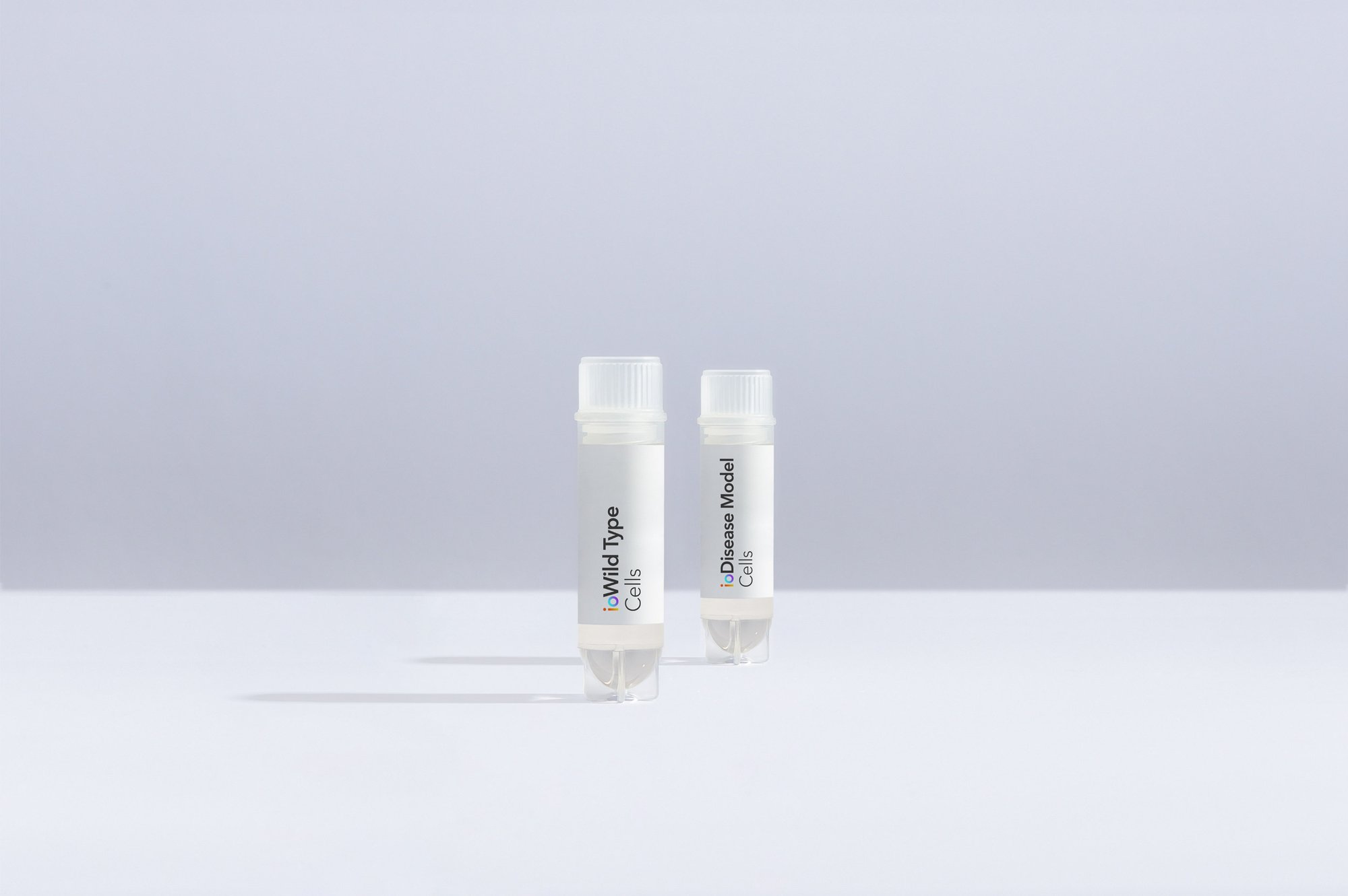
Wild Type and Isogenic Disease Model cells: A true comparison

Further your disease research by pairing our wild type cells with isogenic disease models.

bitbio-vials-Wild_and_Disease-staggered-2500px_wide

ioCells catalogue

Human iPSC-derived cells

powered by opti-ox

Consistent. Defined. Scalable.

bitbio-cell_catalogue_header-with-tracker-Desktop-2500x1664