cat no | io1088
A rapidly maturing, consistent and scalable isogenic system to study Parkinson’s disease (PD).
ioGlutamatergic Neurons SNCA A53T/A53T are opti-ox deterministically programmed glutamatergic neurons carrying a genetically engineered homozygous A53T mutation in the SNCA gene encoding the alpha-synuclein protein.
This disease model is part of a Parkinson’s disease panel of human iPSC-derived cells that can be incorporated into translational research and drug discovery workflows. Two additional clones for the SNCA A53T hom mutation are available for scientists who wish to repeat their experiments in multiple independent clones, please enquire. All disease models are genetically matched to the wild-type control, ioGlutamatergic Neurons. Additional mutations in the PD panel include heterozygous SNCA A53T, het and hom PINK1 Q456X, PRKN R275W and GBA mutations.
Confidently investigate your phenotype of interest across multiple clones with our disease model clone panel. Detailed characterisation data (below) and bulk RNA sequencing data (upon request) help you select specific clones if required.
per vial
A maximum number of 20 vials applies. If you would like to order more than 20 vials, please contact us at orders@bit.bio.
Make True Comparisons
Pair the ioDisease Model Cells with the genetically matched wild-type ioGlutamatergic Neurons to directly investigate the effect of the alpha-synuclein mutation on cellular and molecular mechanisms and cell function.
Scalable
With opti-ox technology, we can make billions of consistently programmed cells, surpassing the demands of industrial workflows.
Quick
The disease model cells and wild-type control are experiment ready as early as 2 days post revival, and form structural neuronal networks at 11 days.
ioGlutamatergic Neurons SNCA A53T/A53T express neuron-specific markers comparably to the wild type control
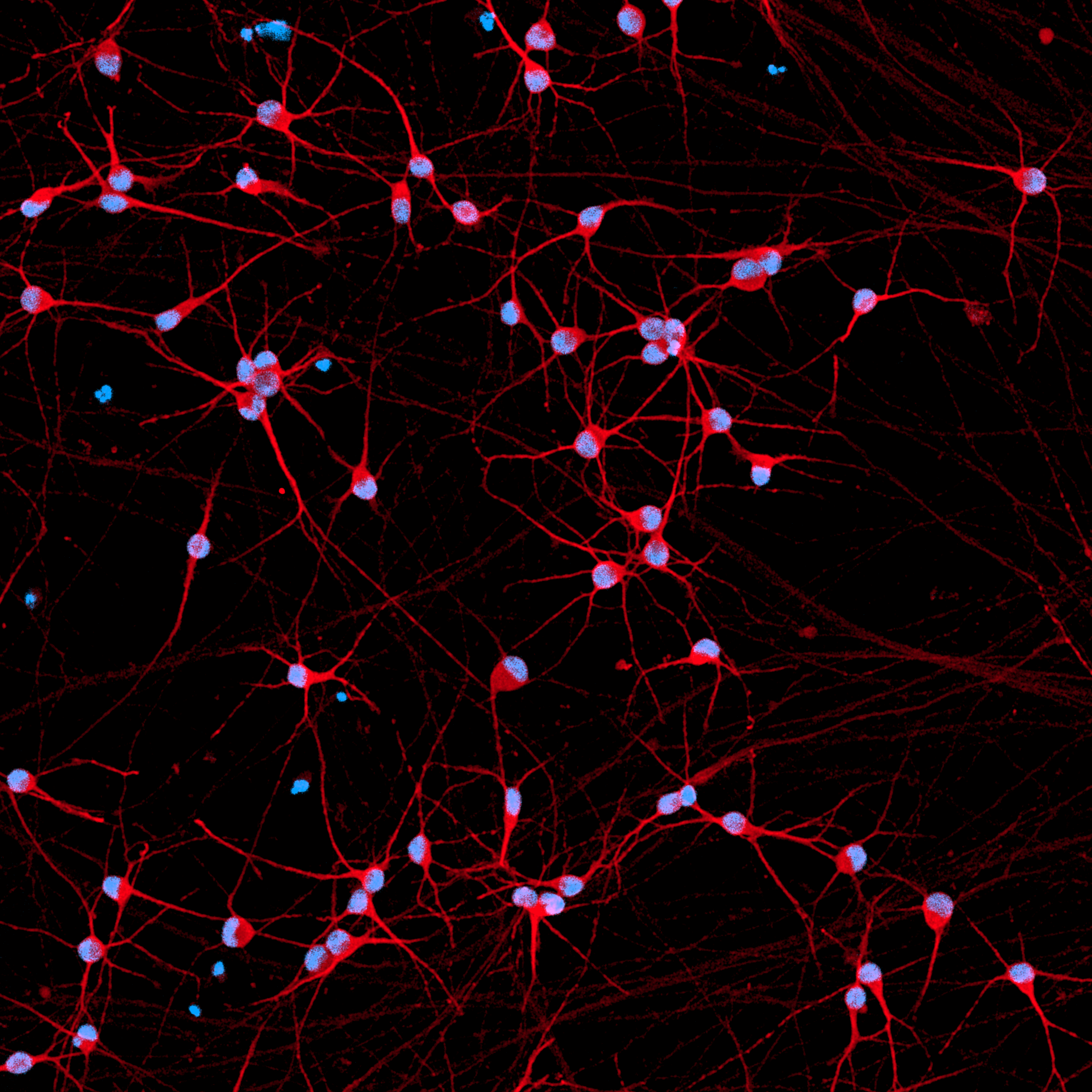
Immunofluorescent staining on day 11 post revival demonstrates similar homogenous expression of pan-neuronal proteins MAP2 and TUBB3 (upper panel) and glutamatergic neuron-specific transporter VGLUT2 (lower panel) in ioGlutamatergic Neurons SNCA A53T/A53T clones compared to the genetically matched control. 100X magnification.
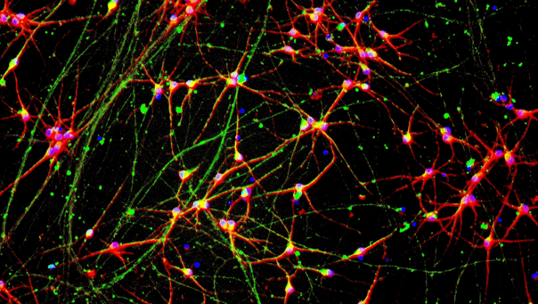
ioGlutamatergic Neurons SNCA A53T/A53T form structural neuronal networks by day 11
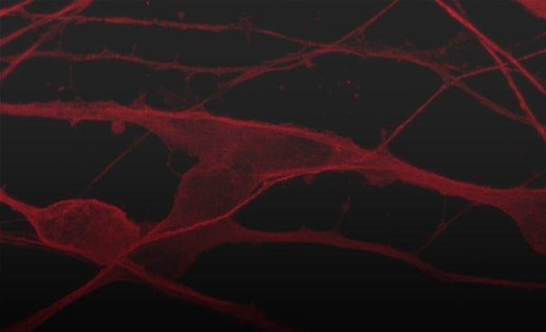
ioGlutamatergic Neurons SNCA A53T/A53T mature rapidly, show glutamatergic neuron morphology and form structural neuronal networks over 11 days, highly similar to the genetically matched control. Day 1 to 11 post thaw; 100X magnification.
ioGlutamatergic Neurons SNCA A53T/A53T demonstrate gene expression of neuronal-specific and glutamatergic-specific markers following deterministic programming
Gene expression analysis demonstrates that ioGlutamatergic Neurons SNCA A53T/A53T and wild-type ioGlutamatergic Neurons (WT Control) lack the expression of pluripotency markers (NANOG and OCT4) at day 11, whilst robustly expressing pan-neuronal (TUBB3 and SYP) and glutamatergic-specific (VGLUT1 and VGLUT2) markers, as well as the glutamate receptor GRIA4. Gene expression levels were assessed by RT-qPCR (data normalised to HMBS; cDNA samples of the parental human iPSC line (hiPSC) were included as reference). Data represents day 11 post-revival samples, n=2 replicates.
Disease-related SNCA is expressed in ioGlutamatergic Neurons SNCA A53T/A53T following deterministic programming
RT-qPCR analysis demonstrates a similar expression level of the SNCA gene in both wild type ioGlutamatergic Neurons (WT Control) and ioGlutamatergic Neurons SNCA A53T/A53T clones at day 11 post-revival (n=2 replicates). cDNA samples of the parental human iPSC line (hiPSC) were included as a reference.
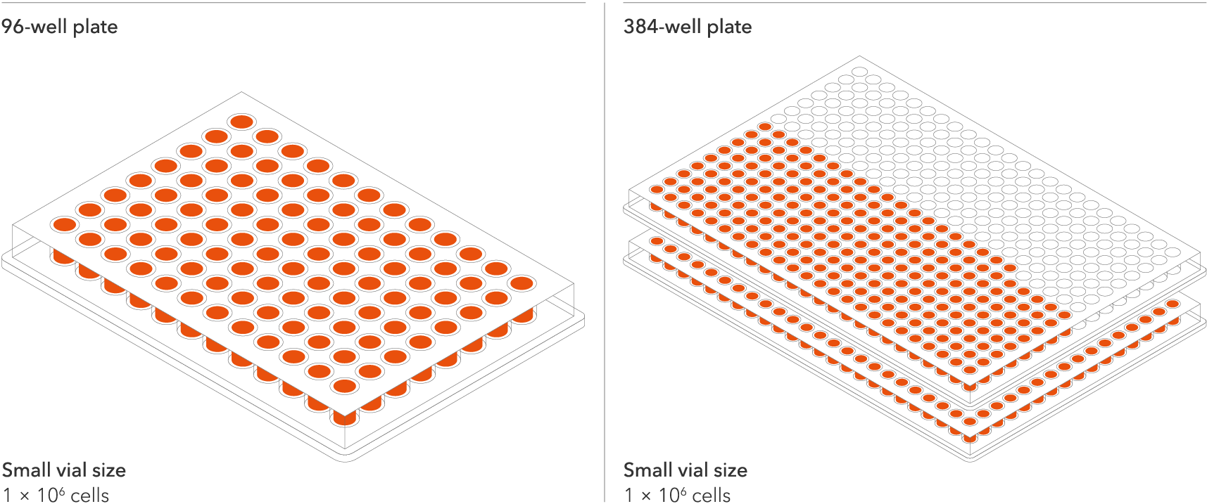
ioGlutamatergic Neurons SNCA A53T/A53T are delivered in a cryopreserved format and are programmed to mature rapidly upon revival in the recommended media. The protocol for the generation of these cells is a two-phase process: Phase 1, Stabilisation for 4 days; Phase 2, Maintenance, during which the neurons mature. Phases 1 and 2 after revival of cells are carried out by the customer.
The recommended minimum seeding density is 30,000 cells/cm2, compared to up to 250,000 cells/cm2 for other similar products on the market. One small vial can plate a minimum of 0.7 x 24-well plate, 1 x 96-well plate, or 1.5 x 384-well plates. This means every vial goes further, enabling more experimental conditions and more repeats, resulting in more confidence in the data.
Starting material
Human iPSC line
Seeding compatibility
6, 12, 24, 96 & 384 well plates
Shipping info
Dry ice
Donor
Caucasian adult male, age 55-60 years old (skin fibroblast)
Vial size
Small: >1 x 10⁶ viable cells
Quality control
Sterility, protein expression (ICC), gene expression (RT-qPCR) and genotype validation (Sanger sequencing)
Differentiation method
opti-ox deterministic cell programming
Recommended seeding density
30,000 cells/cm²
User storage
LN2 or -150°C
Format
Cryopreserved cells
Product use
ioCells are for research use only
Genetic modification
Homozygous A53T missense mutation in the SNCA gene
Applications
Parkinson's disease research
Drug discovery and development
Disease modelling
Available clones
io1087: ioGlutamatergic Neurons SNCA A53T/A53T (D1)
io1088: ioGlutamatergic Neurons SNCA A53T/A53T (H5)
io1089: ioGlutamatergic Neurons SNCA A53T/A53T (H8)
bit.bio
V11
bit.bio
2024
Professor Deepak Srivastava
Professor of Molecular Neuroscience and Group Leader, MRC Centre for Developmental Disorders
King’s College London
Emmanouil Metzakopian | Vice President, Research and Development | bit.bio
Javier Conde-Vancells | Director Product Management | bit.bio
Chakraborty et al
Nature Communications
2023
Featuring ioGlutamatergic Neurons
Dr Ania Wilczynska | Head of Computational Genomics | Non-Clinical | bit.bio
Innovation showcase talk at ISSCR
Marius Wernig MD, PhD | Stanford
Mark Kotter, MD, PhD | bit.bio
Oosterveen, et al
bit.bio & Charles River Laboratories
2023
Qiaojin Lin et al
The EMBO Journal
2023
Featuring opti-ox powered hiPSC-derived glutamatergic neurons with constitutive expression of Cas9
Read this blog on glutamatergic neuron cell culture for our top tips on careful handling, cell plating and media changes to achieve success from the outset.
Further your disease research by pairing our wild type cells with isogenic disease models.