ioGlutamatergic Neurons PRKN R275W/WT
Human iPSC-derived Parkinson's disease model (PRKN mutation ) in human iPSC-derived glutamatergic neurons
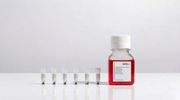
bitbio-ioGlutamatergic-Neurons-media-kits
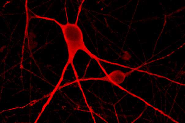
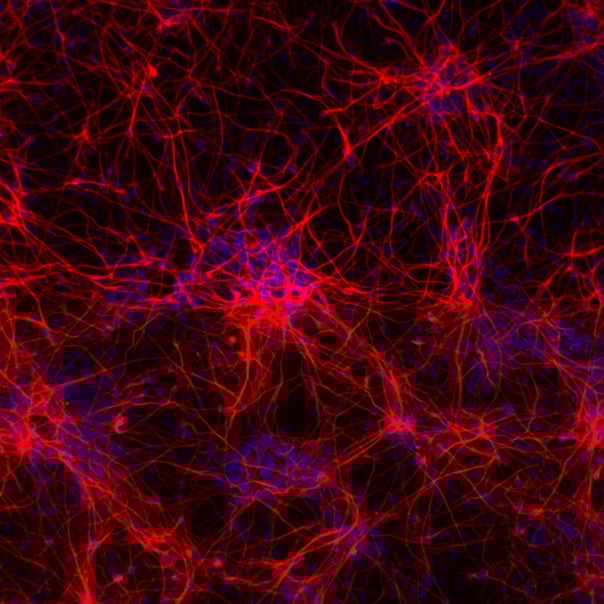
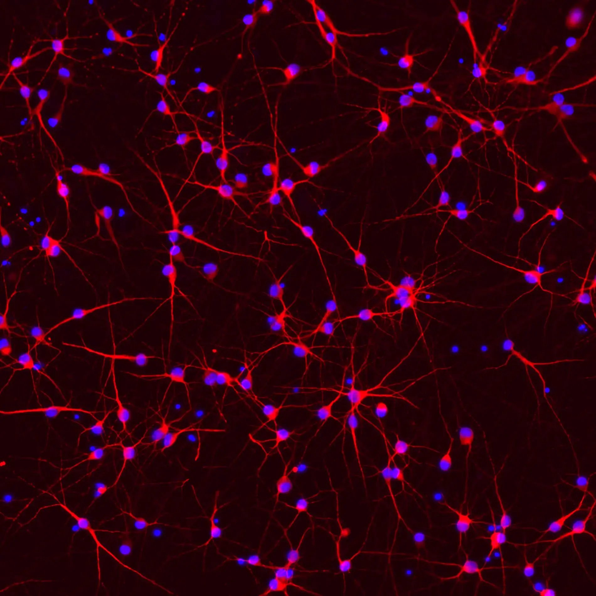
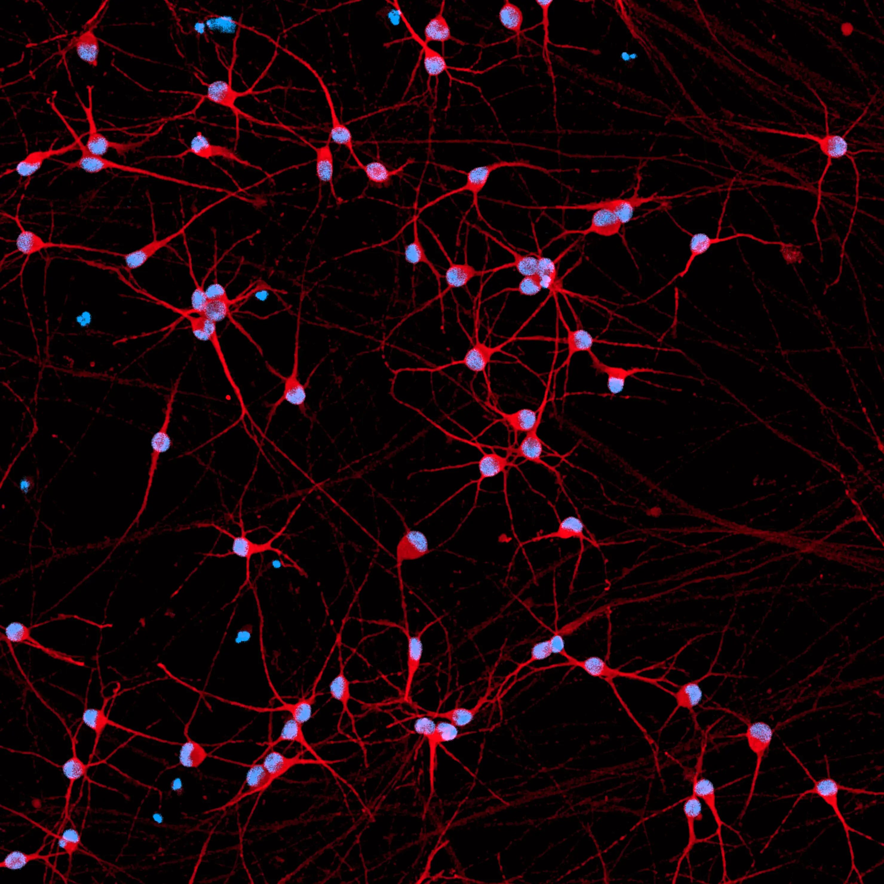
ICC showing glutamatergic neurons with heterozygous R275W mutation in the PRKN gene expressing pan-neuronal markers in an equivalent manner to an isogenic control
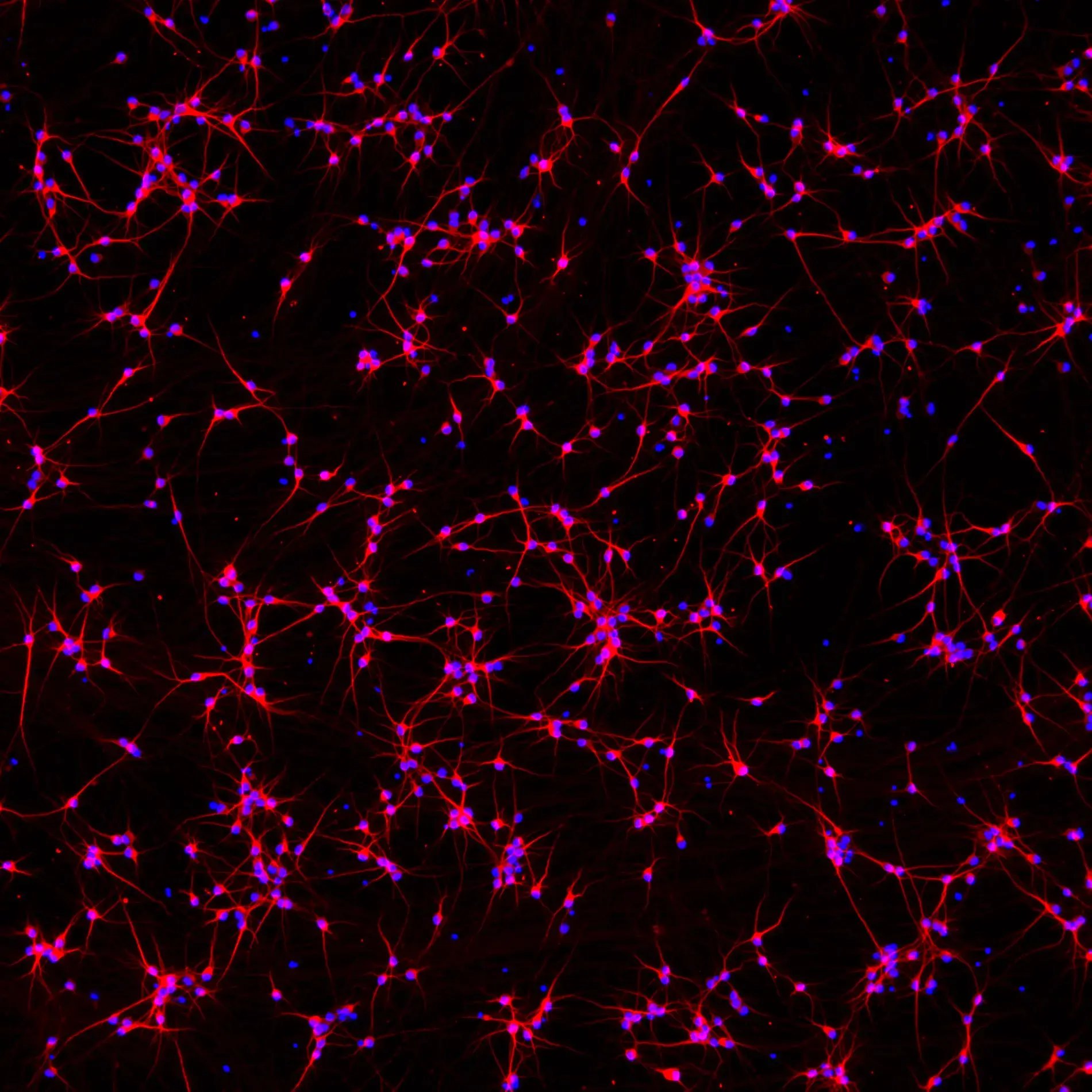
Incucyte imaging of ioGlutamatergic Neurons PRKN R275W/WT over 11 days, showing the cells form structural neuronal networks
RT-qPCR of ioGlutamatergic Neurons PRKN R275W het showing expression of pan neuronal and glutamatergic markers
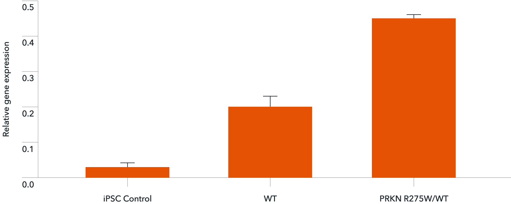
RT-qPCR of ioGlutamatergic Neurons PRKN R275W/WT showing expression of the PRKN gene
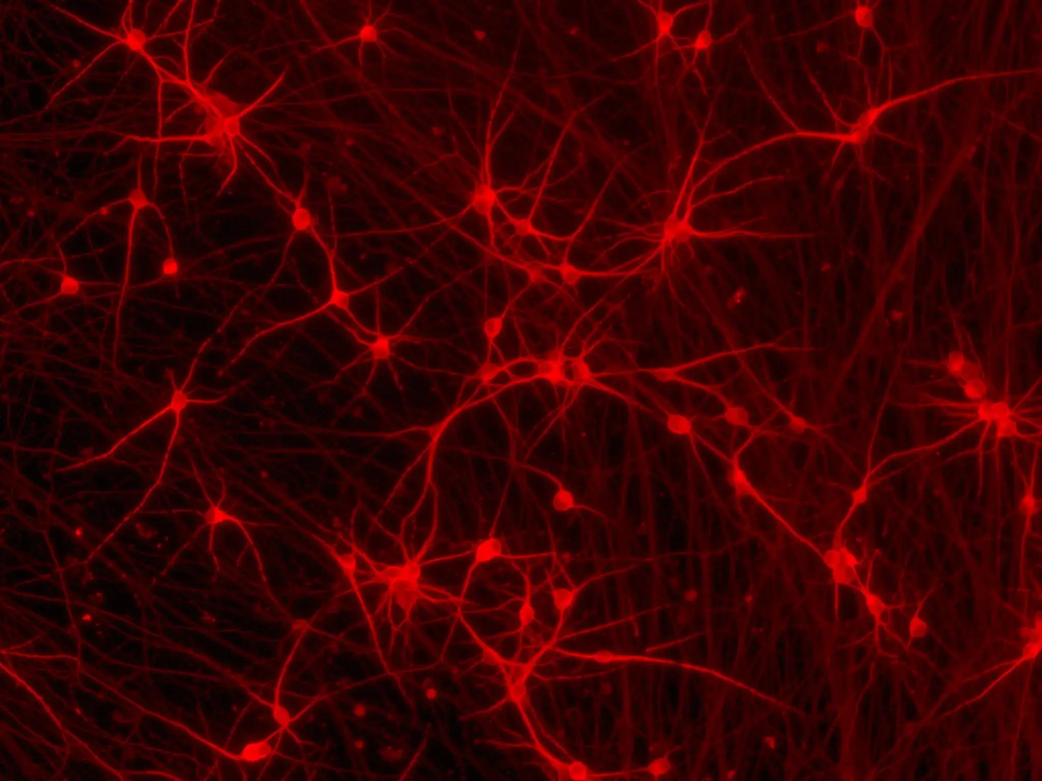
Phenotypic analysis of a tri-culture for CNS modelling using ioGlutamatergic Neurons, ioAstrocytes, and ioMicroglia
mRNA transfection of human iPSC-derived neurons with GFP.
bitbio-lipid-delivery-glutamatergic-neurons-GFP-quantification
Commercially available human iPSC-derived glutamatergic neurons
Human iPSC-derived Parkinson's disease model (PRKN mutation ) in human iPSC-derived glutamatergic neurons
bitbio-ioGlutamatergic-Neurons-media-kits
ICC showing glutamatergic neurons with heterozygous R275W mutation in the PRKN gene expressing pan-neuronal markers in an equivalent manner to an isogenic control
Incucyte imaging of ioGlutamatergic Neurons PRKN R275W/WT over 11 days, showing the cells form structural neuronal networks
RT-qPCR of ioGlutamatergic Neurons PRKN R275W het showing expression of pan neuronal and glutamatergic markers
RT-qPCR of ioGlutamatergic Neurons PRKN R275W/WT showing expression of the PRKN gene
Phenotypic analysis of a tri-culture for CNS modelling using ioGlutamatergic Neurons, ioAstrocytes, and ioMicroglia
mRNA transfection of human iPSC-derived neurons with GFP.
bitbio-lipid-delivery-glutamatergic-neurons-GFP-quantification
Commercially available human iPSC-derived glutamatergic neurons

cat no | io1013

ioGlutamatergic Neurons PRKN R275W/WT

Human iPSC-derived Parkinson's disease model

  • Cryopreserved human iPSC-derived cells powered by opti-ox that are ready for experiments in days
  • In vitro cell model engineered to carry a mutation in PRKN for Parkinson's disease research
  • Consistent, functional excitatory neurons that form neuronal networks within days
Human iPSC-derived Parkinson's disease model (PRKN mutation ) in human iPSC-derived glutamatergic neurons

Human iPSC-derived Parkinson's disease model

bitbio-ioGlutamatergic-Neurons-media-kits

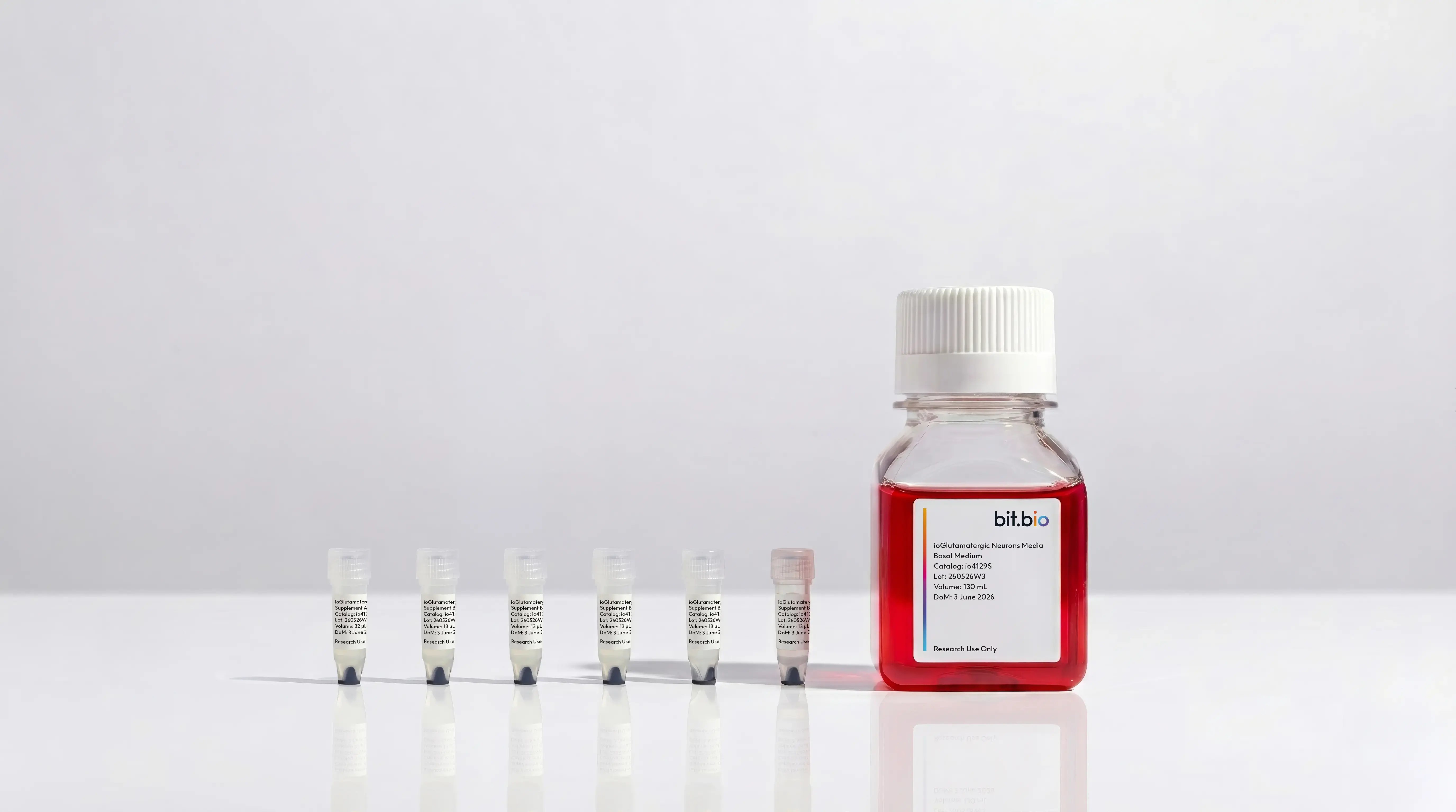
ioGlutamatergic Neurons Media Kit

Cell culture media kit for the culture of ioGlutamatergic Neurons up to 14 days post-thaw

Learn more about the product >

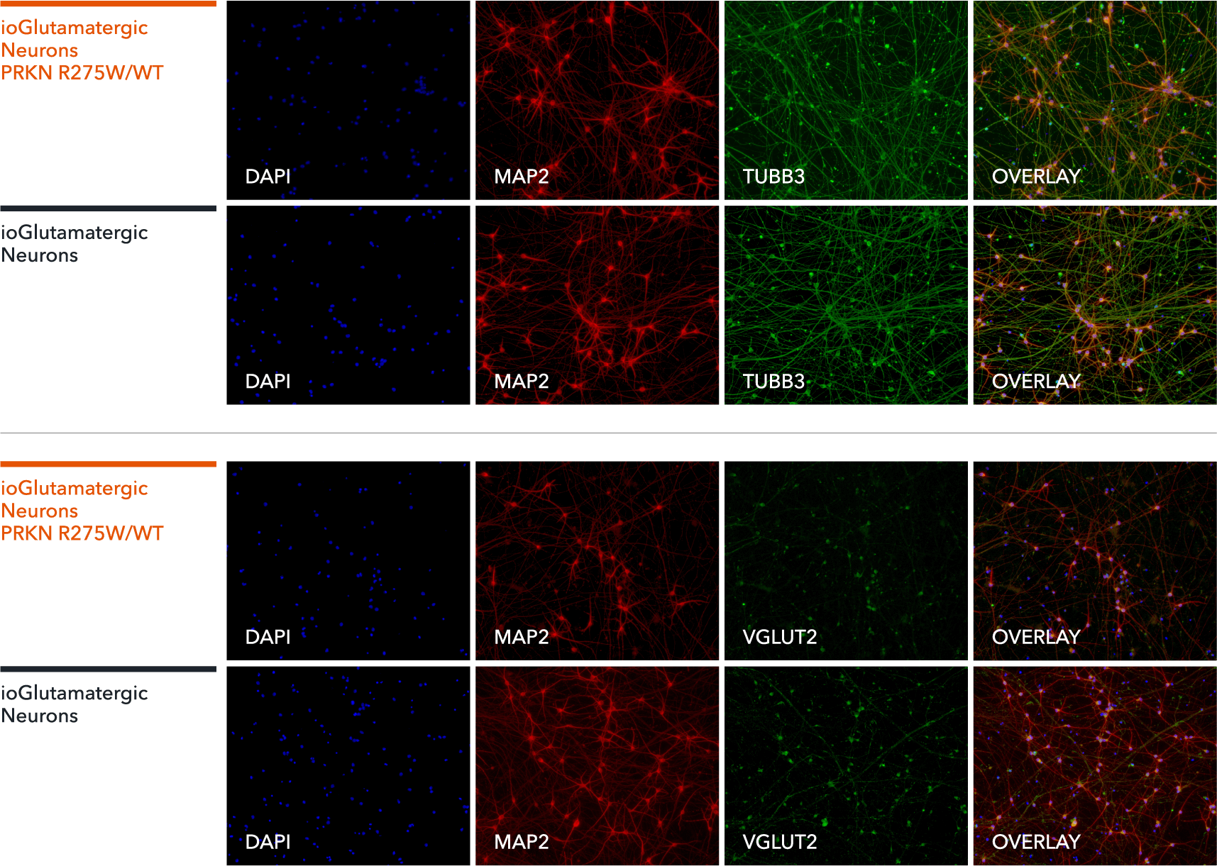
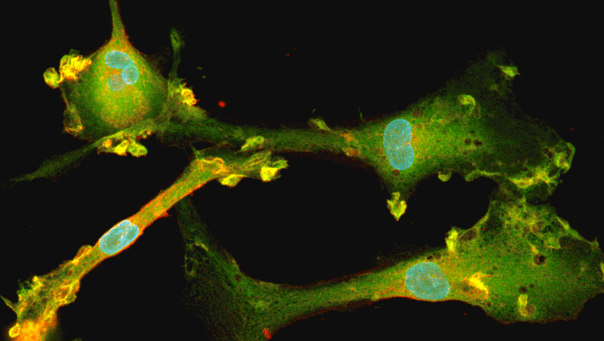
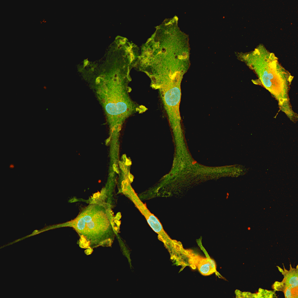
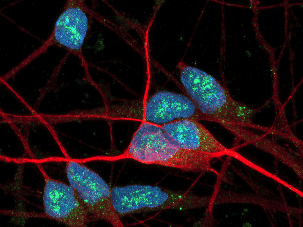
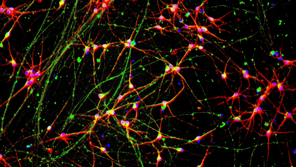
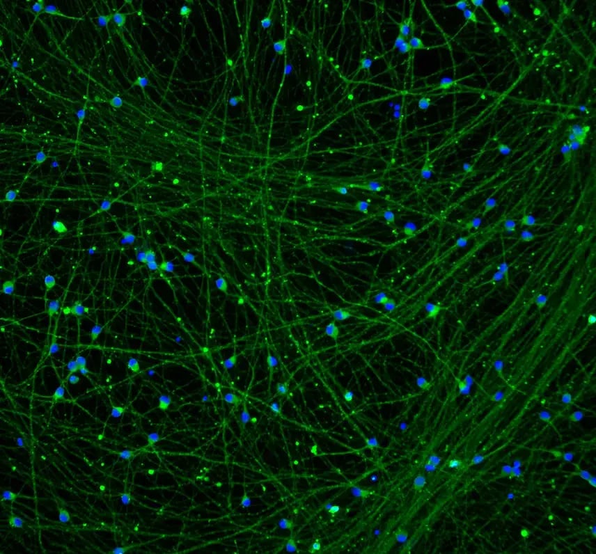
ICC showing glutamatergic neurons with heterozygous R275W mutation in the PRKN gene expressing pan-neuronal markers in an equivalent manner to an isogenic control

ioGlutamatergic Neurons PRKN R275W/WT express neuron-specific markers comparably to the genetically matched control

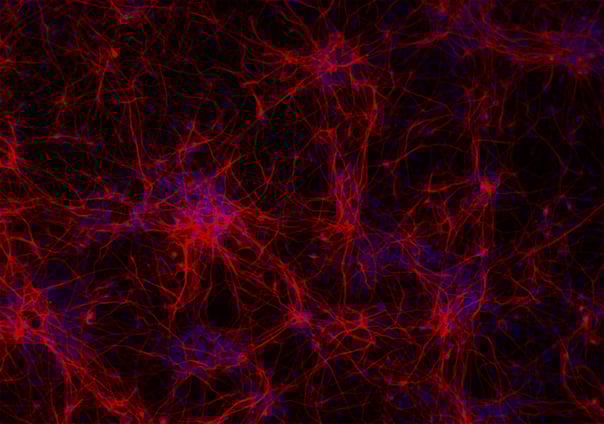
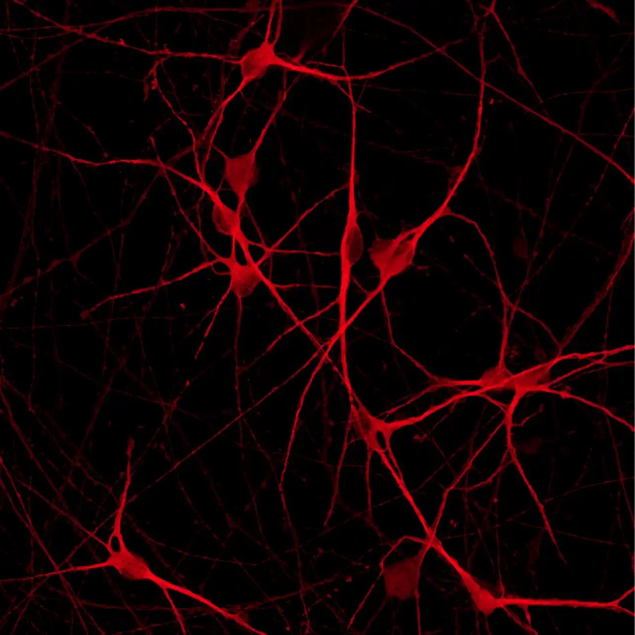
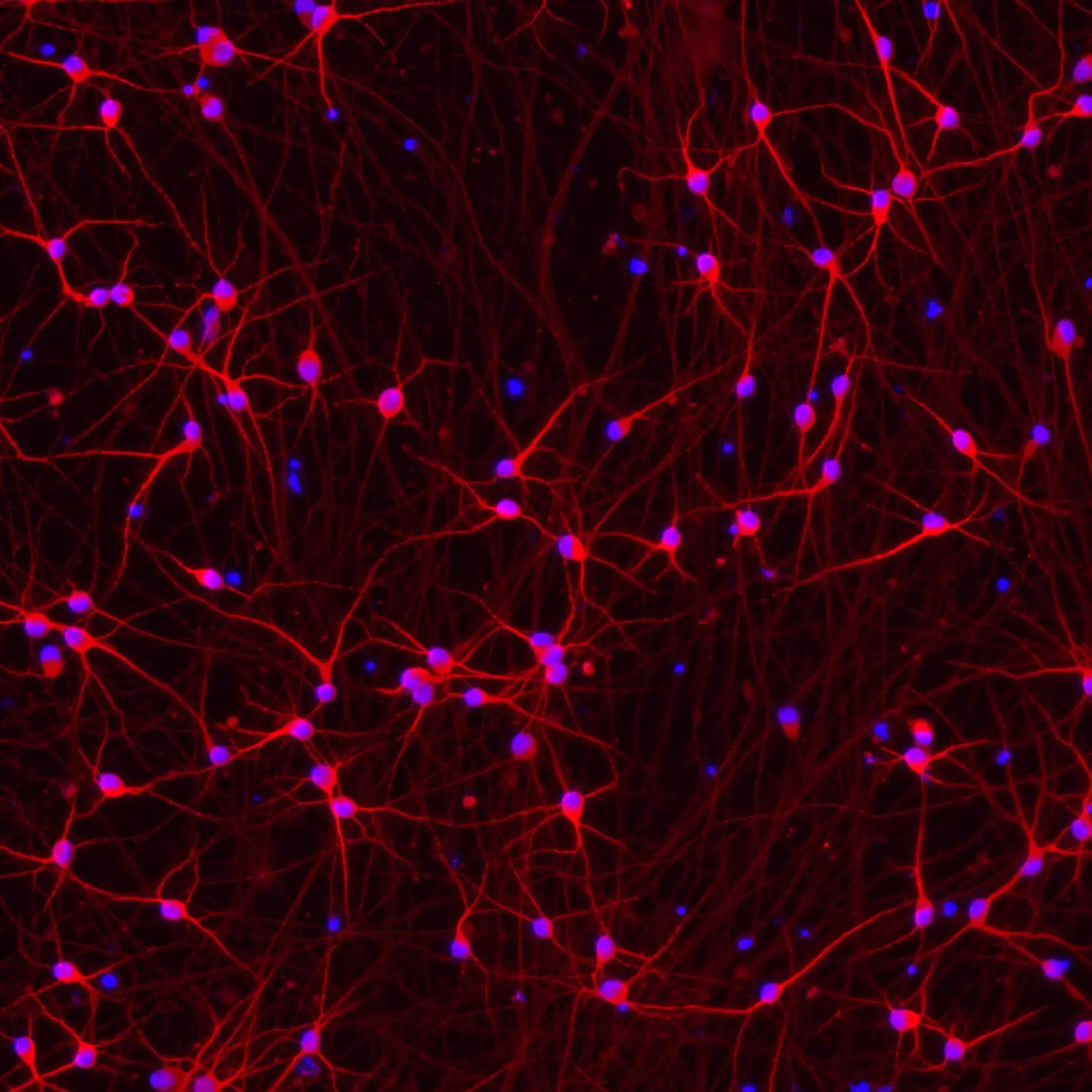
Immunofluorescent staining on post-revival day 11 demonstrates similar homogenous expression of pan-neuronal proteins TUBB3 and MAP2 (upper panel) and glutamatergic neuron-specific transporter VGLUT2 (lower panel) in ioGlutamatergic Neurons PRKN R275W/WT compared to the wild-type control. 100X magnification.

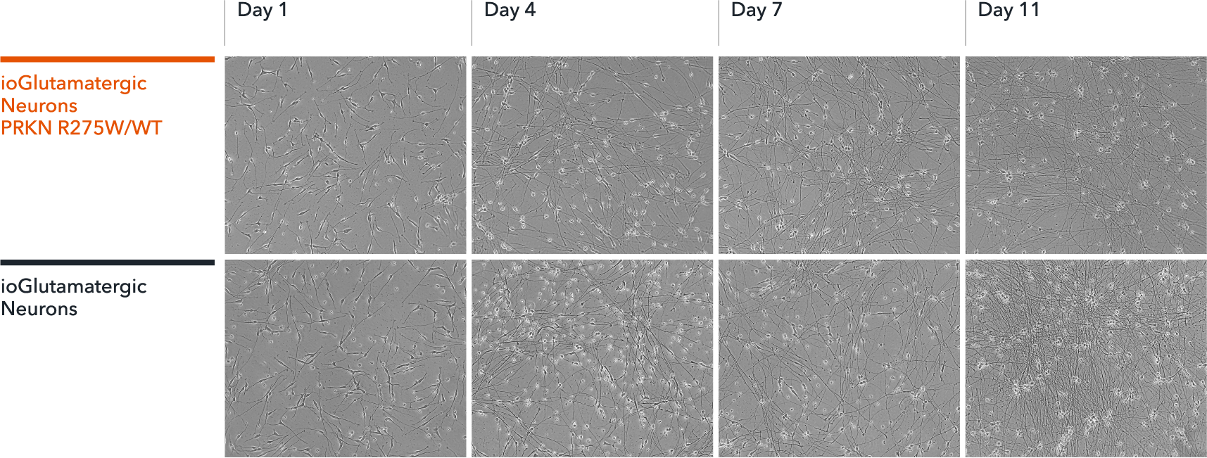
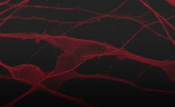
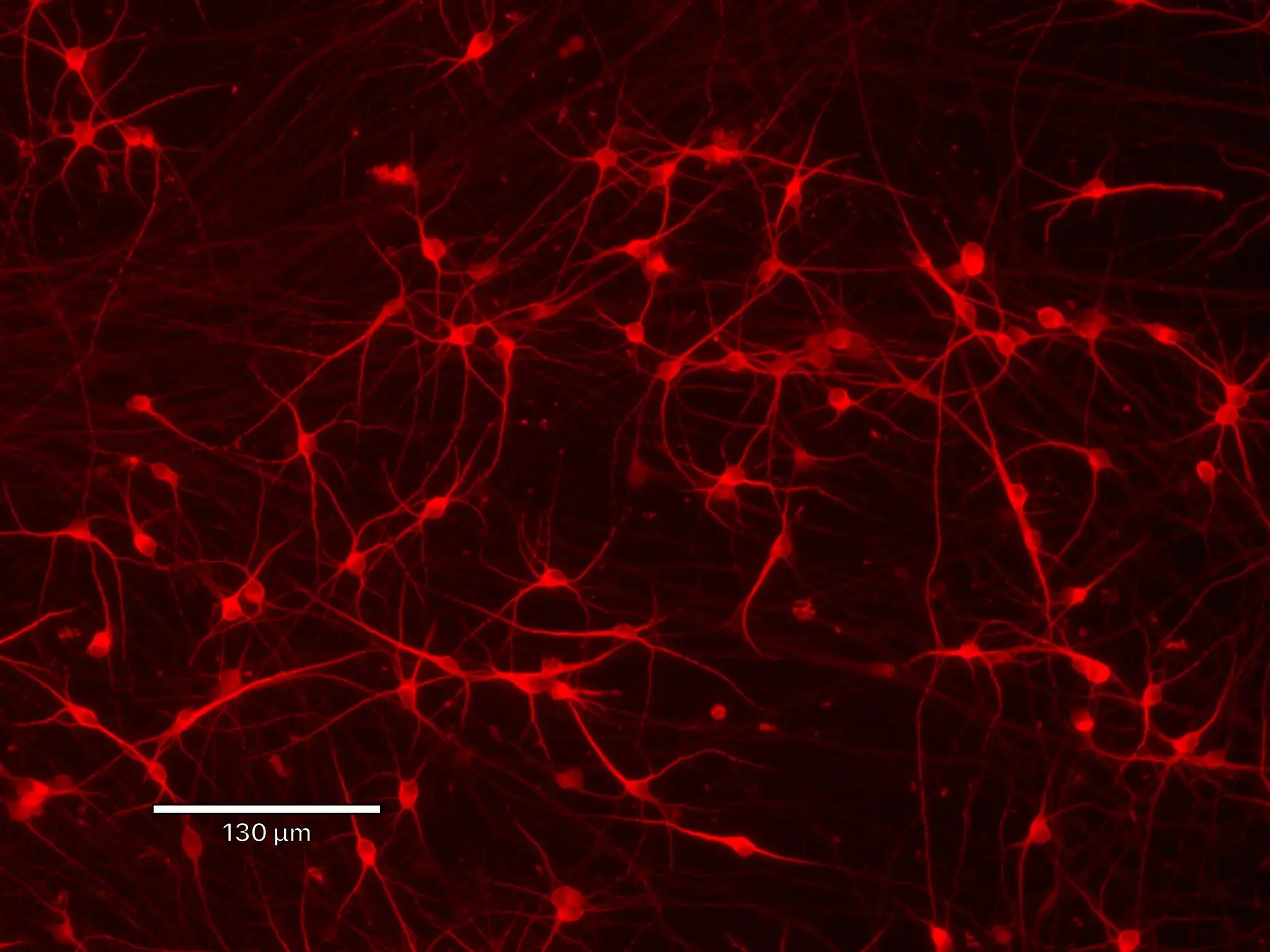
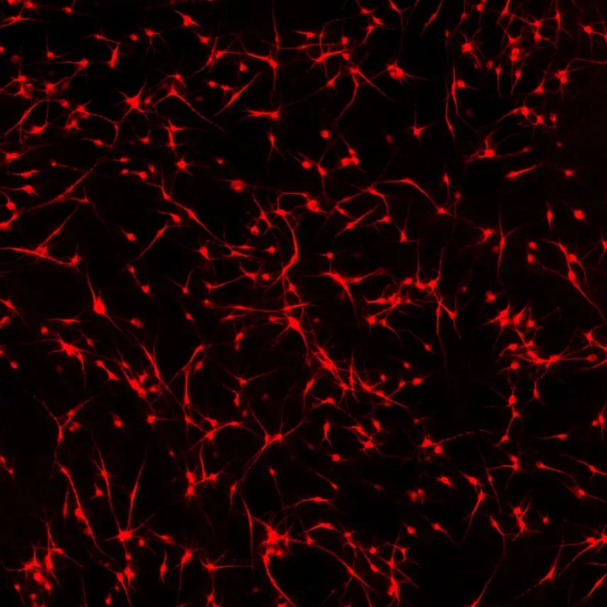
Incucyte imaging of ioGlutamatergic Neurons PRKN R275W/WT over 11 days, showing the cells form structural neuronal networks

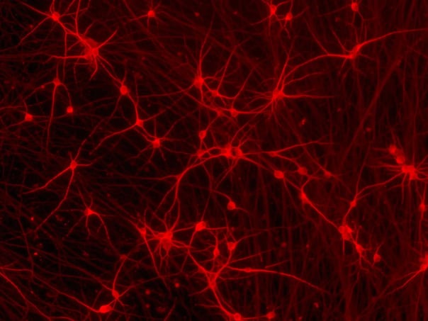
ioGlutamatergic Neurons PRKN R275W/WT form structural neuronal networks by day 11

ioGlutamatergic Neurons PRKN R275W/WT mature rapidly, show glutamatergic neuron morphology and form structural neuronal networks over 11 days, when compared to the wild-type control. Day 1 to 11 post thawing; 100X magnification.

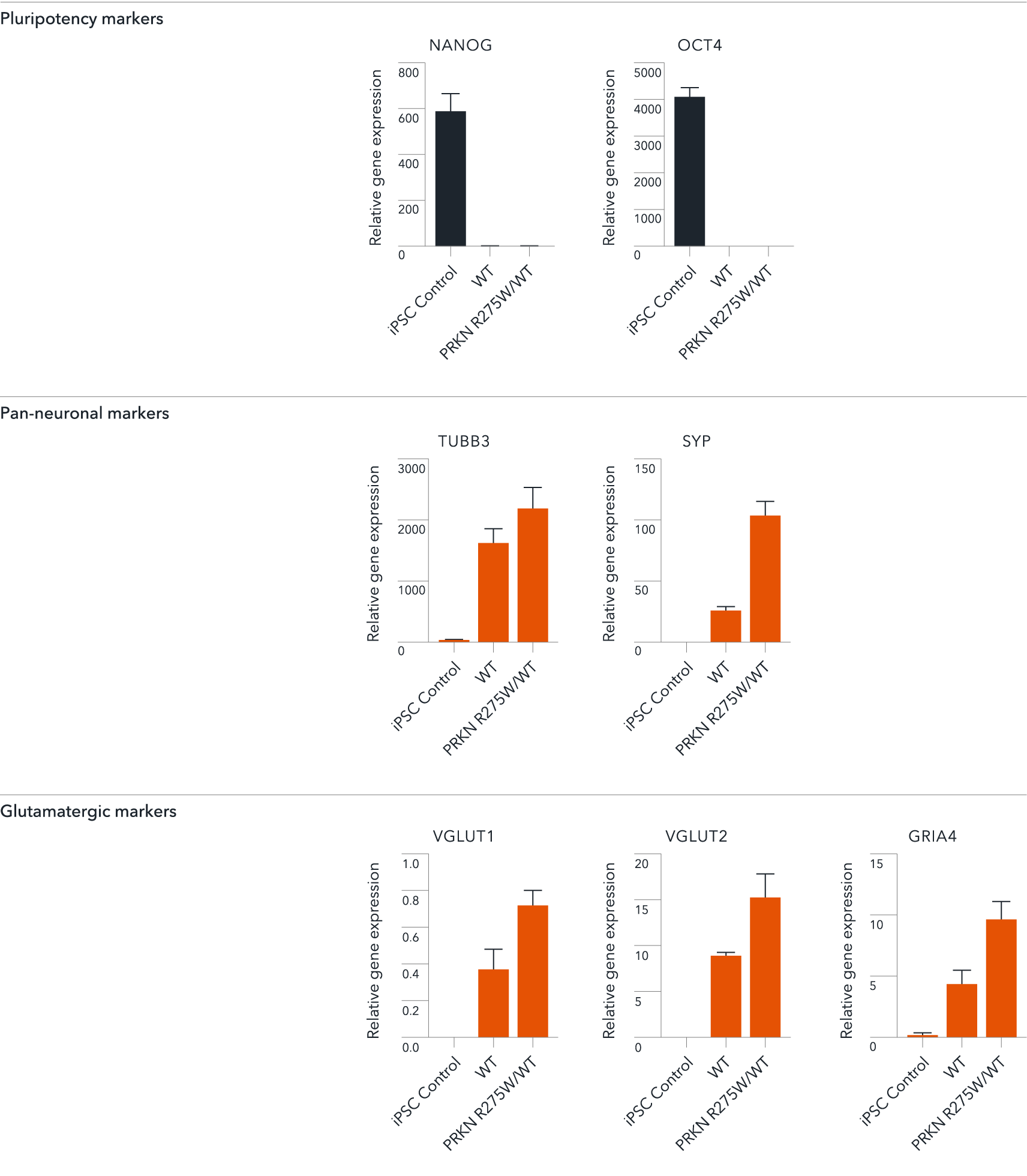
RT-qPCR of ioGlutamatergic Neurons PRKN R275W het showing expression of pan neuronal and glutamatergic markers

ioGlutamatergic Neurons PRKN R275W/WT demonstrate gene expression of neuronal and glutamatergic-specific markers following deterministic programming

Gene expression analysis demonstrates that ioGlutamatergic Neurons PRKN R275W/WT and the wild-type control (WT) lack the expression of pluripotency markers (NANOG and OCT4) at day 11, whilst robustly expressing pan-neuronal (TUBB3 and SYP) and glutamatergic specific (VGLUT1 and VGLUT2) markers, as well as the glutamate receptor GRIA4. Gene expression levels were assessed by RT-qPCR (data normalised to HMBS; cDNA samples of the parental human iPSC line (hiPSC) were included as reference). Data represents day 11 post-revival samples, n=2 replicates.

View the step-by-step RNA extraction and RT-qPCR protocol used to generate this data 

RT-qPCR of ioGlutamatergic Neurons PRKN R275W/WT showing expression of the PRKN gene

Disease-related PRKN is expressed in ioGlutamatergic Neurons PRKN R275W/WT following deterministic programming

RT-qPCR analysis demonstrates expression of the PRKN gene in both wild type ioGlutamatergic Neurons (WT) and ioGlutamatergic Neurons PRKN R275W/WT at day 11 post revival. Data normalised to HMBS, n=2 replicates.

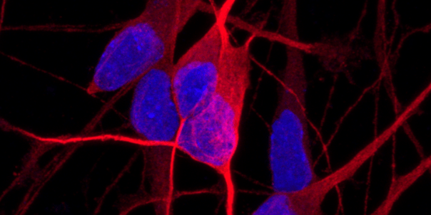
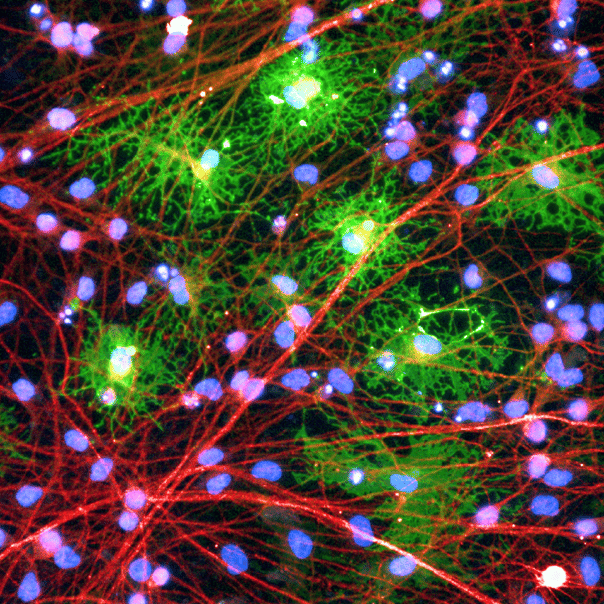
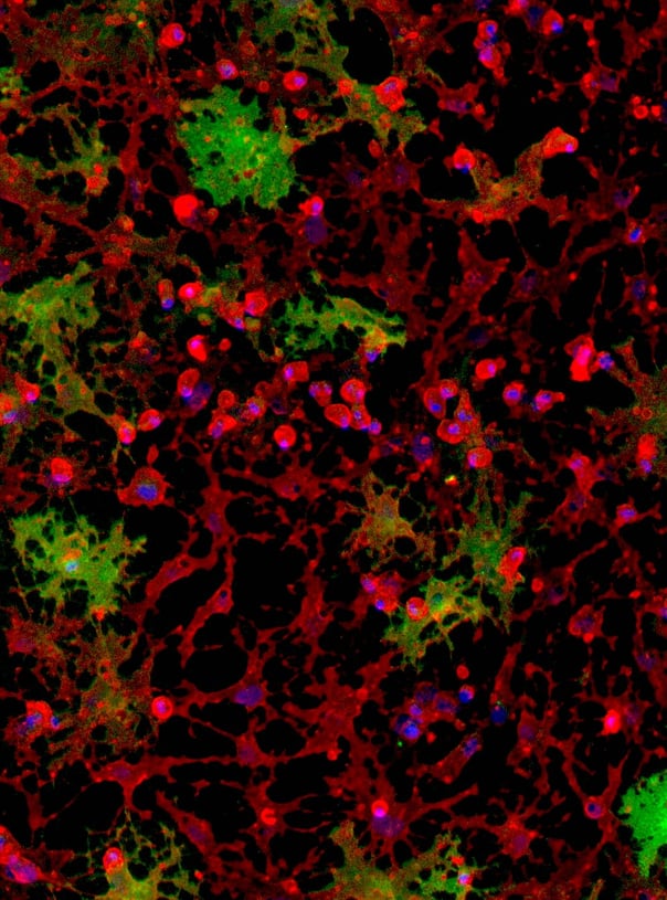
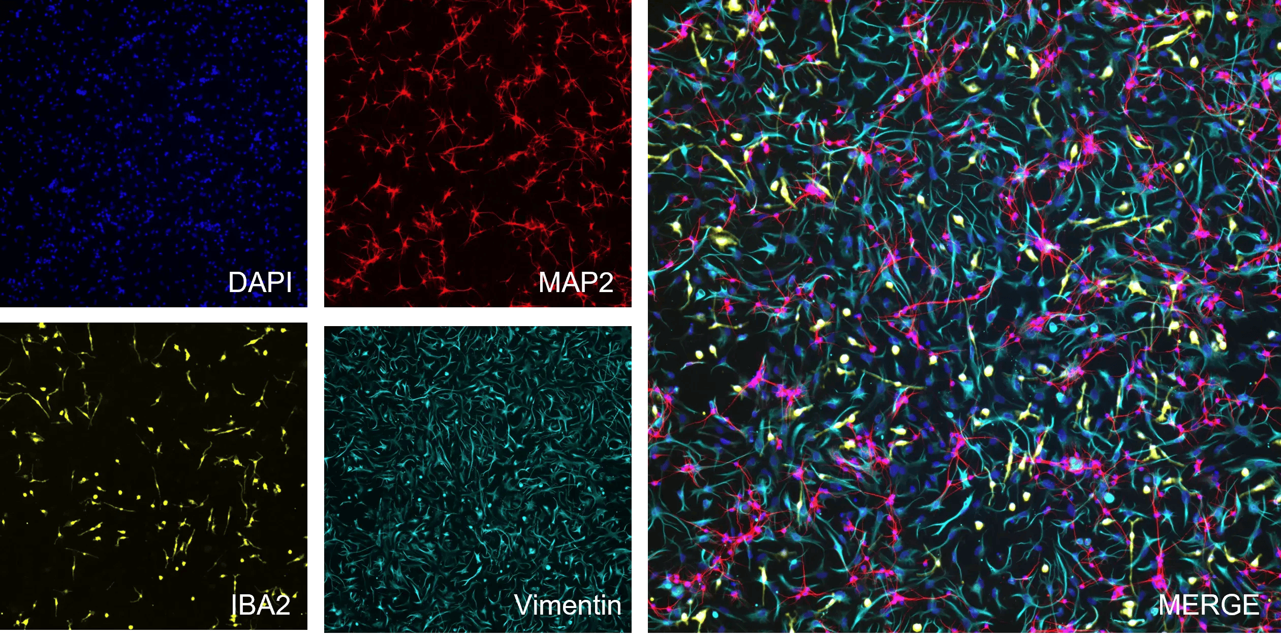
Phenotypic analysis of a tri-culture for CNS modelling using ioGlutamatergic Neurons, ioAstrocytes, and ioMicroglia

Phenotypic characterisation of a human iPSC-derived tri-culture using ioGlutamatergic Neurons, ioAstrocytes, and ioMicroglia

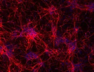
Using our fully optimised protocol, ioGlutamatergic Neurons (MAP2, red), ioMicroglia (IBA1, yellow) and ioAstrocytes (vimentin, cyan) were co-cultured to create a highly defined CNS model. High-resolution ICC analysis confirms the successful co-localisation and morphological health of three distinct cell types within a unified environment. By day 7, the protocol yields a highly consistent, integrated network suitable for complex cell modelling. DAPI (blue) highlights the total cell density and integrity of the culture. This protocol is compatible with derivative products of the three cell types, ensuring straightforward implementation across experimental workflows.

Download the optimised tri-culture protocol

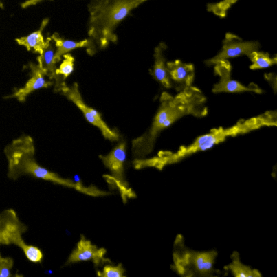
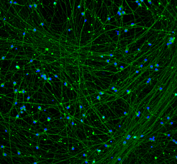
mRNA transfection of human iPSC-derived neurons with GFP.

Efficient mRNA transfection into ioGlutamatergic Neurons

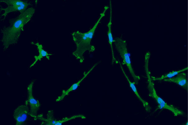
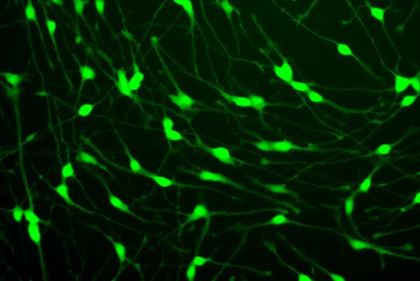
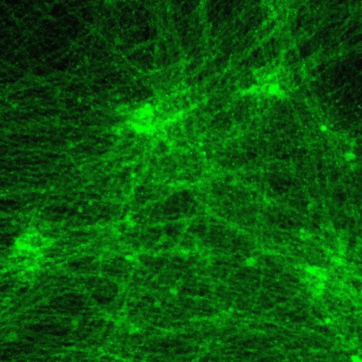
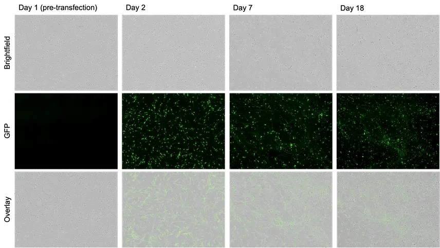
ioGlutamatergic Neurons are efficiently transfected and show sustained long-term expression of mRNA encoding GFP. ioGlutamatergic Neurons were imaged from day 1 post-thaw and throughout the experiment to assess transfection efficiency and evaluate potential cytotoxic effects of the transfection protocol. Day 1 images were captured prior to transfection on the same day.

Download the step-by-step protocol for lipid-based delivery of synthetic mRNA into ioGlutamatergic Neurons.

bitbio-lipid-delivery-glutamatergic-neurons-GFP-quantification

Lipid-based delivery of synthetic mRNA into ioGlutamatergic Neurons

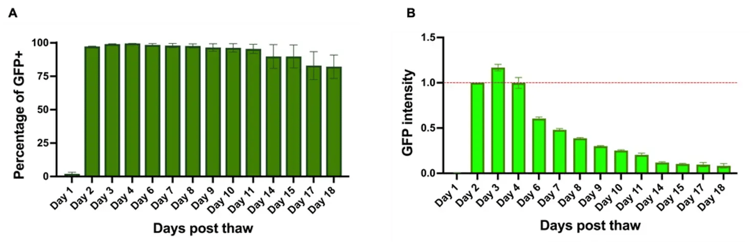
ioGlutamatergic Neurons were transfected 24 hours post-thaw using Lipofectamine™ Stem Transfection Reagent. The transfection efficiency was evaluated by fluorescence imaging over 18 days after mRNA delivery, resulting in high transfection efficiency (close to 100%) and long-term sustained GFP expression.

Quantification of the GFP signal shows a decrease in GFP intensity over time, while the percentage of GFP-positive cells remains largely unchanged over time.

(A) The percentage of GFP-positive cells from two independent experiments.

(B) GFP intensity, quantified in successfully transfected cells from two independent experiments is quantified and normalised to day 2 (24 hours post-transfection).

Explore the full dataset and complete protocol for lipid-based delivery of synthetic mRNA in 96-well plates

Commercially available human iPSC-derived glutamatergic neurons

Industry leading seeding density

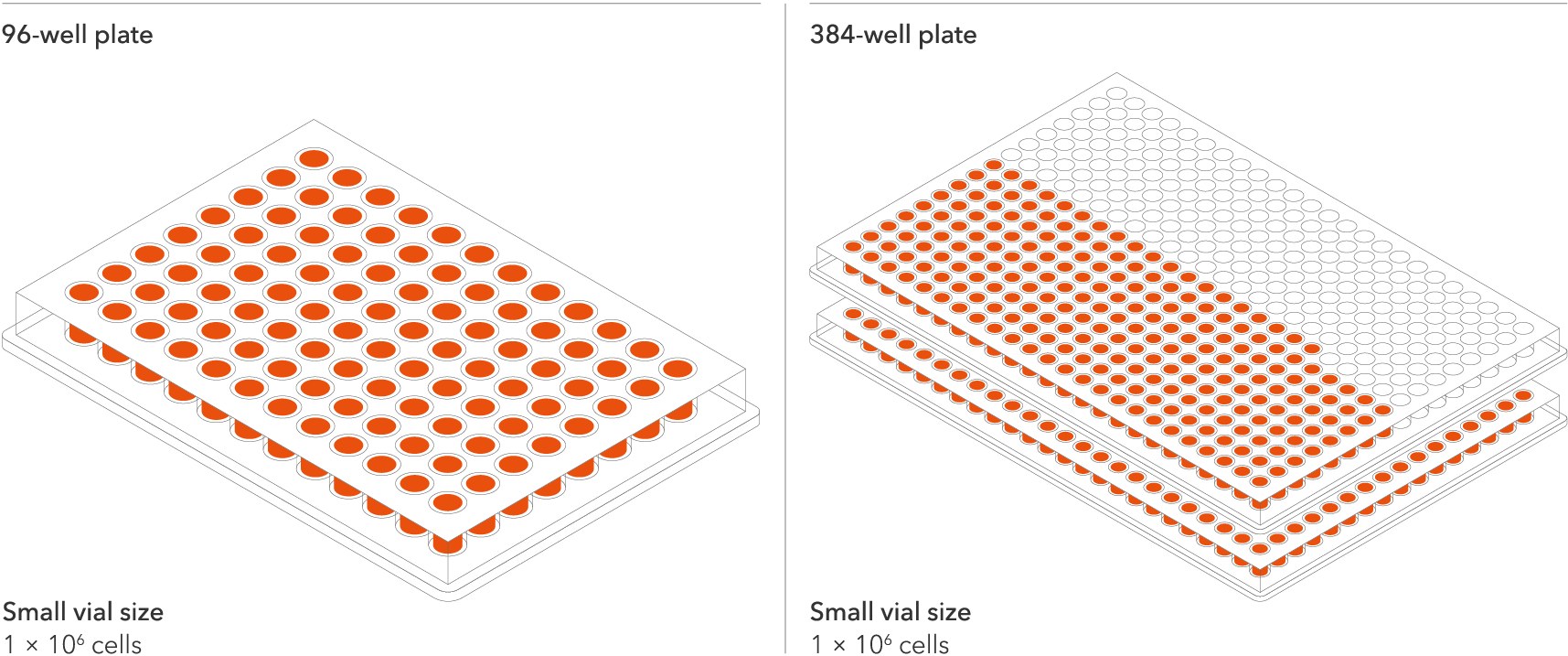
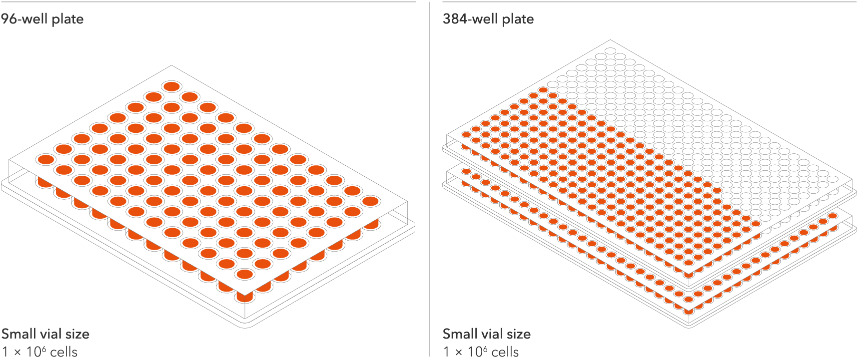
The recommended minimum seeding density is 30,000 cells/cm2, compared to up to 250,000 cells/cm2 for other similar products on the market. One small vial can plate a minimum of 0.7 x 24-well plate, 1 x 96-well plate, or 1.5 x 384-well plates. This means every vial goes further, enabling more experimental conditions and more repeats, resulting in more confidence in the data.

Vial limit exceeded

A maximum number of 20 vials applies. If you would like to order more than 20 vials, please contact us at orders@bit.bio.


Human iPSC-derived Parkinson's disease model

ioGlutamatergic Neurons PRKN R275W/WT are opti‑ox deterministically programmed excitatory neurons carrying a genetically engineered heterozygous mutation in the PRKN gene encoding the Parkin protein. These cells offer a rapidly maturing, disease relevant and isogenic system for investigating the molecular and cellular significance of a heterozygous R275W mutation in Parkinson’s disease.

This disease model is part of a Parkinson's disease panel of physiologically relevant human iPSC-derived cells that can be incorporated into translational research and drug discovery workflows. Additional mutations in the PD panel include a homozygous PRKN R275W mutation, homozygous and heterozygous PINK1 Q456X, SNCA A53T and GBA mutations. All can be used alongside their genetically matched control, ioGlutamatergic Neurons.

Benchtop benefits

comparison_0

Make True Comparisons

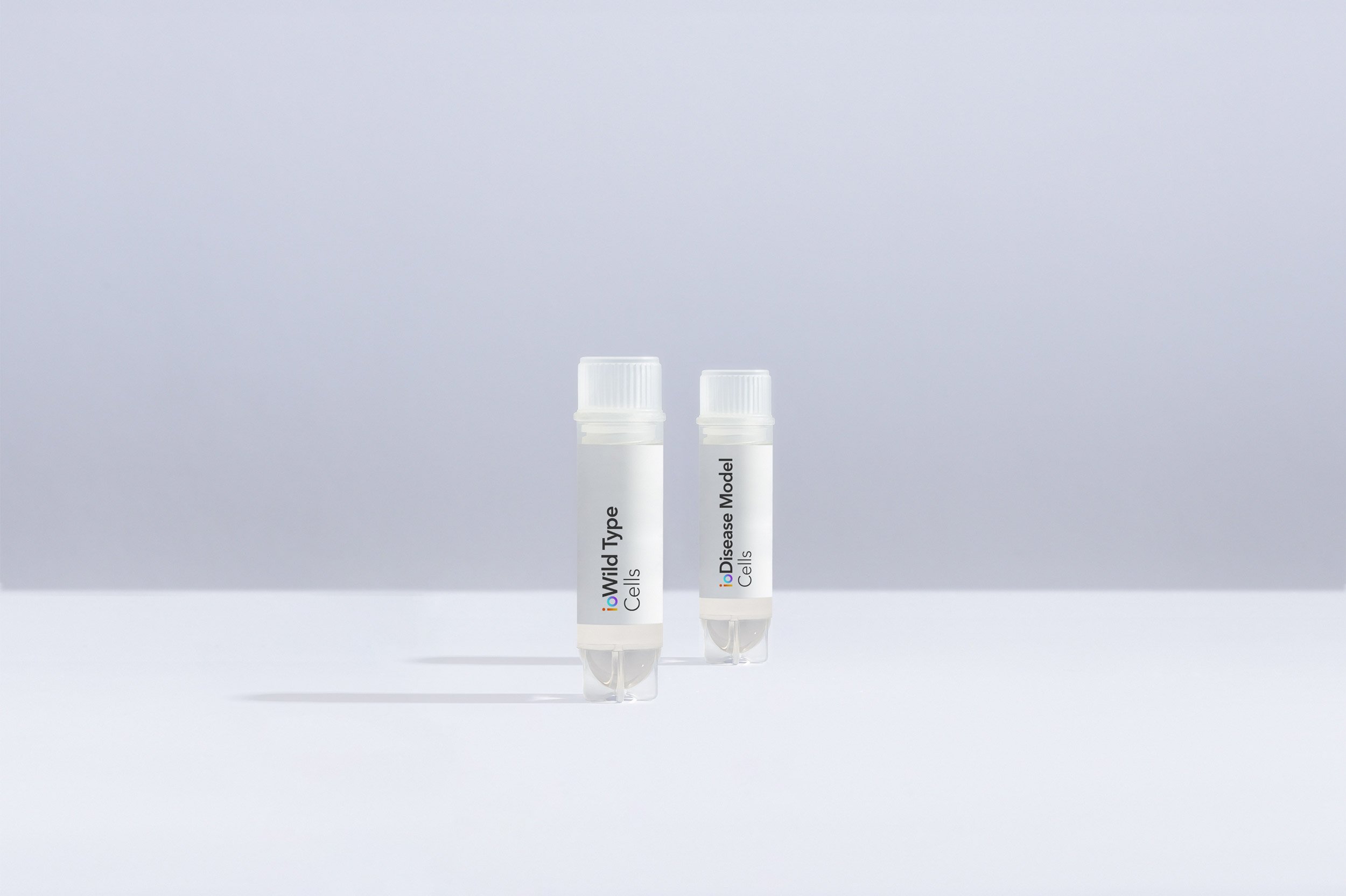
Pair the ioDisease Model Cells with the genetically matched wild-type ioGlutamatergic Neurons to directly investigate the effect of heterozygous expression of mutant Parkin protein on disease.

scalable_0

Scalable

With opti-ox technology, we can make billions of consistently programmed cells, surpassing the demands of industrial workflows.

quick_0

Quick

The disease model cells and isogenic control are experiment ready as early as 2 days post revival, and form structural neuronal networks at 11 days.

Cells arrive ready to plate


ioGlutamatergic_Neurons_and_disease_models_timeline

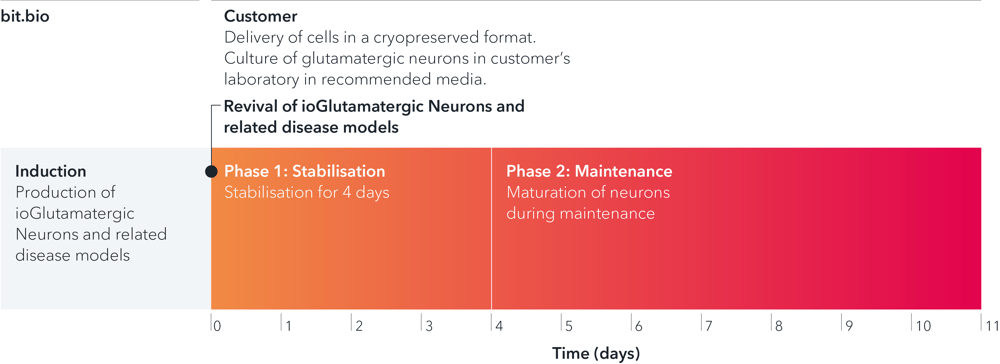
ioGlutamatergic Neurons PRKN R275W/WT are delivered in a cryopreserved format and are programmed to mature rapidly upon revival in the recommended media. The protocol for the generation of these cells is a two-phase process: Phase 1, Stabilisation for 4 days; Phase 2, Maintenance, during which the neurons mature. Phases 1 and 2 after revival of cells are carried out by the customer.

Product specifications

Starting material

Human iPSC line

Karyotype

Normal (46, XY)

Seeding compatibility

6, 12, 24, 48, 96 & 384 well plates

Shipping info

Dry ice

Donor

Caucasian adult male, age 55-60 years old (skin fibroblast),
Genotype APOE 3/4

Vial size

Small: >1 x 10⁶ viable cells

Quality control

Sterility, protein expression (ICC), gene expression (RT-qPCR) and genotype validation (Sanger sequencing)

Differentiation method

opti-ox deterministic cell programming

Recommended seeding density

30,000 cells/cm2

User storage

LN2 or -150°C

Format

Cryopreserved cells

Genetic modification

Heterozygous R275W missense mutation in the PRKN gene

Applications

Parkinson's disease research
Drug discovery and development
Disease modelling

Product use

ioCells are for research use only


Scale your study with volume pricing

Enabling scientists to use human cells in their research, running additional experiments without rationing cells or limiting experimental scale


Order quantity Total vials received Pricing tier 
1 - 9 packs 3 - 27 vials Standard price
10 - 33 packs 30 - 99 vials Automatic 10% discount
> 34 packs > 100 vials > Contact us for a quote


Academic pricing: Academic users can purchase any ioCells in 3-vial packs ($/€/£ 999 per pack), available year-round with any cell type combination.
Technical data

Highly characterised and defined

ioGlutamatergic Neurons PRKN R275W/WT express neuron-specific markers comparably to the genetically matched control

ICC showing glutamatergic neurons with heterozygous R275W mutation in the PRKN gene expressing pan-neuronal markers in an equivalent manner to an isogenic control

Immunofluorescent staining on post-revival day 11 demonstrates similar homogenous expression of pan-neuronal proteins TUBB3 and MAP2 (upper panel) and glutamatergic neuron-specific transporter VGLUT2 (lower panel) in ioGlutamatergic Neurons PRKN R275W/WT compared to the wild-type control. 100X magnification.

ioGlutamatergic Neurons PRKN R275W/WT form structural neuronal networks by day 11

Incucyte imaging of ioGlutamatergic Neurons PRKN R275W/WT over 11 days, showing the cells form structural neuronal networks

ioGlutamatergic Neurons PRKN R275W/WT mature rapidly, show glutamatergic neuron morphology and form structural neuronal networks over 11 days, when compared to the wild-type control. Day 1 to 11 post thawing; 100X magnification.

ioGlutamatergic Neurons PRKN R275W/WT demonstrate gene expression of neuronal and glutamatergic-specific markers following deterministic programming

RT-qPCR of ioGlutamatergic Neurons PRKN R275W hom showing expression of pan neuronal and glutamatergic markers

Gene expression analysis demonstrates that ioGlutamatergic Neurons PRKN R275W/WT and the wild-type control (WT) lack the expression of pluripotency markers (NANOG and OCT4) at day 11, whilst robustly expressing pan-neuronal (TUBB3 and SYP) and glutamatergic specific (VGLUT1 and VGLUT2) markers, as well as the glutamate receptor GRIA4. Gene expression levels were assessed by RT-qPCR (data normalised to HMBS; cDNA samples of the parental human iPSC line (hiPSC) were included as reference). Data represents day 11 post-revival samples, n=2 replicates.

View the step-by-step RNA extraction and RT-qPCR protocol used to generate this data 

Disease-related PRKN is expressed in ioGlutamatergic Neurons PRKN R275W/WT following deterministic programming

RT-qPCR of ioGlutamatergic Neurons PRKN R275W/WT showing expression of the PRKN gene

RT-qPCR analysis demonstrates expression of the PRKN gene in both wild type ioGlutamatergic Neurons (WT) and ioGlutamatergic Neurons PRKN R275W/WT at day 11 post revival. Data normalised to HMBS, n=2 replicates.

Industry leading seeding density

ioGlutamatergic_Neurons_seeding_density_small_96_384

The recommended minimum seeding density is 30,000 cells/cm2, compared to up to 250,000 cells/cm2 for other similar products on the market. One small vial can plate a minimum of 0.7 x 24-well plate, 1 x 96-well plate, or 1.5 x 384-well plates. This means every vial goes further, enabling more experimental conditions and more repeats, resulting in more confidence in the data.

How to culture ioGlutamatergic Neurons

In this video, our scientist will take you through the step-by-step process of how to thaw, seed and culture ioGlutamatergic Neurons.

How to prepare tissue culture vessels with PDL-Geltrex for culture

Dr Kaiser Karim will take you through the step-by-step process of coating your culture plate with PDL-Geltrex before the revival of ioGlutamatergic Neurons.

Since recording this video Geltrex has been replaced by Geltrex Flex; refer to the user manual for full details. 

Frequently Asked Questions (FAQs)


  • Why is the R275W point mutation in the PRKN gene relevant for disease modelling?

Mutations in the Parkin gene are associated with the second most common genetic form of Parkinson's Disease and are known to cause mitochondrial dysfunction and neuroinflammation.


  • Is there a genetically-matched wild-type control that can be used alongside the PRKN disease model in human iPSC-derived glutamatergic neurons?

bit.bio offers the PRKN R275W heterozygous and homozygous disease models and a genetically-matched wild-type control, which provide a physiologically-relevant model to investigate the impact of PRKN R275W mutation on the cellular and molecular mechanisms and function in Parkinson's Disease.

Product resources

Cell culture hacks | human iPSC-derived glutamatergic neurons 

Read this blog on glutamatergic neuron cell culture for our top tips on careful handling, cell plating and media changes to achieve success from the outset.

bit.bio_3x2_ioGlutamatergic Neurons_MAP2_Hoescht_x20_hi.res (1)

Wild Type and Isogenic Disease Model cells: A true comparison.

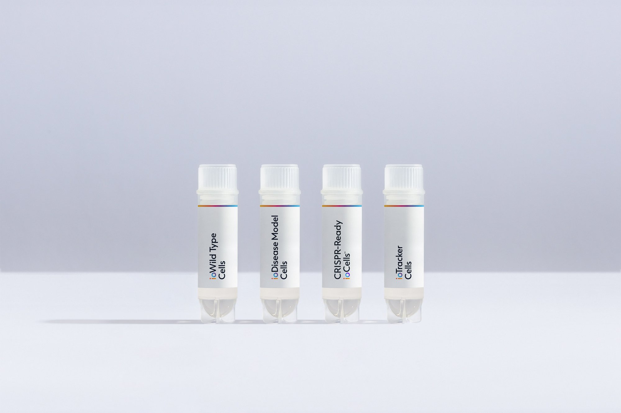
Further your disease research by pairing our wild type cells with isogenic disease models.

bitbio-vials-Wild_and_Disease-staggered-2500px_wide-B

ioCells catalogue

Human iPSC-derived cells

powered by opti-ox

Consistent. Defined. Scalable.

bitbio-cell_catalogue_header-with-tracker-Desktop-2500x1664